

 PDF
PDF ePub
ePub Citation
 Print
Print


INTRODUCTION
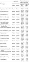
Opitz syndrome (OS; MIM 145410 and 300000) is also known as hypertelorism-hypospadias syndrome, and it is a congenital midline malformation syndrome that was formerly reported as two separate entities, G and BBB syndromes (1, 2). The constellation of clinical manifestations that define OS include: 1) congenital heart defects such as atrial and ventricular septal defects, patent ductus arteriosus and coarctation of the aorta; 2) a characteristic facial appearance (a broad nasal bridge, hypertelorism and low-set posteriorly rotated ears) with labiopalatine and laryngotracheal abnormalities; 3) dysphagia and gastro-oesophageal reflux; 4) abnormalities of the central nervous system (including major motor skill defects and delayed development); and 5) genital anomalies, e.g. hypospadias (3).
OS is genetically heterogeneous because it has both an X-linked (Xp22.3) and an autosomal (22q11.2) loci (4). The gene responsible for X-linked OS (XLOS) is the MID1 gene which encodes a microtubule-associated RING-Bbox-Coiled-coil (RBCC) protein (5). Mutations in the MID1 gene have been found either in familial or sporadic cases, and indicated as the cause of the X-linked form of OS (XLOS). To date, about 35 distinct MID1 mutations including missense, nonsense, and splicing mutations, small insertions, small deletions and complex rearrangements have been identified and most of those are concentrated in the 3' end of the gene (5-7). We recently found a four-year male patient with multiple midline defects and we identified a novel mutation in the MID1 gene in this patient and his mother, which enabled us to make a successful prenatal diagnosis for two consecutive fetuses.
MATERIALS AND METHODS
Patients
The male patient was born in 2002 by Cesarean section after 40 weeks of pregnancy and he was the first live born baby. The birth weight was approximately 3,800 g and he was grunting immediately after delivery. He presented with hyperterolism, a broad nasal bridge, a laryngeal cleft and hypospadias. He experienced many episodes of aspiration pneumonia and gastroesophageal reflux. His mother has hypertelorism without any other dysmorphic findings (Table 1).
Fluorescent in situ hybridization (FISH)
To rule out the autosomal form of OS, FISH studies using the TUPLE1 probe for the DiGeroge/VCFS critical region (Vysis, Downers Grove, IL, U.S.A.) were accomplished according to the manufacturer's specifications with minor modifications. We analyzed 200 interphase nuclei from peripheral blood lymphocytes of the patient.
Mutation analysis
Genomic DNA was extracted from peripheral blood leukocytes with using the Wizard Genomic DNA Purification kit (Promega, Madison, WI, U.S.A.) according to the manufacturer's instructions. All the coding exons and their flanking introns of the MID1 gene were amplified by using primer sets that were designed by the authors (these are available upon request). The polymerase chain reaction (PCR) was performed with a thermal cycler (Model 9700, Applied Biosystems, Foster City, CA, U.S.A.) as follows: 32 cycles of denaturation at 94℃ for 30 sec, annealing at 60℃ for 30 sec and extension at 72℃ for 30 sec. After treating the amplicon (5 µL) with 10 U shrimp alkaline phosphatase and 2 U exonuclease I (USB Corp., Cleveland, OH, U.S.A.), direct sequencing was performed with the BigDye Terminator Cycle Sequencing Ready Reaction kit (Applied Biosystems) on an ABI Prism 3100 genetic analyzer (Applied Biosystems).
Prenatal diagnosis
After identifying the causative mutation in this family, chorionic villus sampling (CVS) was performed at 74 days of gestation in a 33-yr-old patient's mother (gravida 2, para 1). The DNA was extracted according to standard procedures, and exon 13 of the MID1 gene was sequenced and analyzed on an ABI Prism 3100 genetic analyzer (Applied Biosystems). Cytogenetic analysis was then done by GTG banding. Amniocentesis was carried out for the next pregnancy and the same procedure was done for the CVS.
RESULTS
Mutation analysis
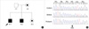
On the FISH analysis, all the cells had two red signals of a similar intensity for TUPLE1 and two green signals of the locus-specific identifier (LSI) ARSA. Mutation analysis of the MID1 gene by direct sequencing of all nine coding exons and intron/exon boundaries identified the c.1798_1799insC mutation in exon 13 (Fig. 1). This mutation is located in the second codon position of the amino acid histidine 600, which produces a frameshift leading to the production of a chimeric 599-residue MID1 protein ending with 12 amino acids that are derived from a wrong reading frame. The patient's mother was a carrier of the same mutation.
Prenatal diagnosis
After identifying the causative mutation in this family, the parents of the patient requested prenatal diagnosis for the next two consecutive fetuses. Genomic DNA was isolated from the chorionic villi of the 2nd baby and from the amniotic fluid of the 3rd baby, and this was subsequently screened for the presence of the c.1798_1799insC mutation by performing sequence analysis of the MID1 exon 13. The mutant-type allele was observed in both fetuses. According to the cytogenetic analysis, the 2nd baby was male and this fetus was terminated according to parents' demand; the 3rd baby was female and delivered.
DISCUSSION
In the present study, we report the results of the MID1 gene mutation analysis that was done in a male patient with XLOS and his mother. After identification of the causative mutation in this family, we successfully performed prenatal diagnosis for the next two fetuses. Our patient had typical phenotypes except for his neurologic abnormalities and congenital heart defects, as compared to those previously reported (5-9). There is considerable clinical variability in the presentation of OS, even among the male patients from the same family who share identical MID1 mutations (7), and this suggests that other factors modifying the OS phenotype may exist. As far as the genotype/phenotype correlations are concerned, Pinson et al. (10) reported that vermis hypoplasia or agenesis was the most common brain anomaly observed in OS patients with MID1 mutations, and this has particularly been seen in association with the R495X mutation (p<0.0001). So et al. (9) suggested that only laryngo-tracheo-esophageal malformations might correlate with MID1 mutation. Recently, Mnayer et al. (11) suggested that OS patients with mutations in the FNIII domain of MID1 resulting retained activity of the FNIII domain would have a milder phenotype without severe brain damage.
Considering the absence of a cure and complete penetrance, prenatal diagnosis appears to be a considerable approach to reduce the burden of this disease on the individual family and also on society. Prenatal diagnosis of OS, including discriminating the sex, is important because the daughters of known or suspected carriers are not likely to be functionally impaired or have an obvious diagnosis of carriers. The finding of hypertelorism as a pervasive sign in the carriers of XLOS, along with a personal or family history of cleft lip, cleft palate or other manifestations of XLOS, may be important as a prenatal screening tool (9). Most XLOS families carry a unique mutation and screening of the whole MID1 gene is laborious and time consuming. Therefore, prenatal direct mutation analysis can be timely done in those families of which the mutation is known beforehand.
In summary, we found a novel MID1 mutation in a Korean male patient suffering from XLOS and his mother, and this enabled us to make a successful prenatal diagnosis in this family. To the best of our knowledge, this is the first report on a genetically confirmed case of XLOS in Korea.
 XML Download