

 PDF
PDF ePub
ePub Citation
 Print
Print


INTRODUCTION
Thyroxine is one of counter-regulatory hormone in glucose homeostasis. Therefore, metabolic control in diabetes is negatively affected by thyrotoxicosis. Typically, hyperglycemic hyperosmolar state (HHS) occurs in elderly individuals with type 2 diabetes mellitus (DM) accompanying with a several week history of polyuria, weight loss, diminished oral intake and decreased mentality in severe cases. HHS is often precipitated by a serious, concurrent illness such as myocardial infarction or systemic infections (1).
Although there have been several case reports describing that diabetic ketoacidosis is associated with thyrotoxicosis in type 2 diabetics(2-4), only one case of HHS associated with Graves' hyperthyroidism developed in type 2 diabetics has been reported in the non-English literature until now (5). We present the case of a young woman with type 2 DM and hyperthyroid Graves' disease developing to HHS.
CASE REPORT
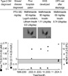
A 23-yr-old woman with type 2 DM and Graves' disease has revisited to the Department of Emergency of Gyeongsang National University Hospital due to nausea, generalized weakness, polyuria and weight loss of 5 kg for 7 days in October 2003 since she was discharged from our hospital three weeks before. She was previously diagnosed as type 2 DM in 1996, and recently started insulin treatment to improve glycemic control due to persistently elevated HbA1c irrespective of oral administration of hypoglycemic agents. Three years before, she had taken propylthiouracil for hyperthyroid Graves' disease for 2 yr. In May 2003, she was diagnosed as relapse of hyperthyroidism at a local clinic and referred to our hospital for management of thyrotoxicosis refractory to usual dosage of propylthiouracil, 300 mg/day and proliferative diabetic retinopathy and nephropathy. She had 7.9 mM/L fasting blood glucose, 1.07 nM/L C-peptide (reference range [RR], 0.1-0.83), 222 mM/L fructosamine (RR, 205-285), 8.3% HbA1c (RR, 4.5-5.6) and 0.01 mU/L TSH and 2.02 pM/L free T3 on thryoid function tests. At that time, Her weight was 54 kg and height was 147 cm (body mass index, 25.0 kg/m2). Her thyroid had the volume of about 40 g and it looked diffusely enlarged and rubbery consistency on palpation. The technetium-99m (Tc-99m) thyroid scan revealed marginally increased and somewhat irregular uptake (3.8%). As from hospitalization, the patient had oral administration of relatively high dose of methimazole (60 mg daily), propranolol (160 mg daily) and 12 drops of Lugol's solution (three times a day) for 14 days, and consecutively lithium carbonate (900 mg daily) for 14 days because of persisting tachycardia (>110/ min), dyspnea on exertion and still high concentration of free T3 (4.44 pM/L). She was discharged with receiving methimazole (60 mg daily) and beta-blocker (Fig. 1).
She had no family history of thyroid disease, but both parents have been suffering from type 2 DM. She denied ingestion of any drugs including alcohol or excessive consumption of fizzy drinks these days. She also has been adhering to administration of antithyroid drug, beta-blocker and insulin (0.5 units/kg/day) since being discharged. On physical examination, the patient had 143/89 mmHg blood pressure, 36.8℃ body temperature, 22/min respiration rate, and 121/min pulse rate. She looked acutely ill, and had slightly tremulous, warmhands, and decreased skin turgor and dried tongue. She was 48 kg weight and body mass index 22.2 kg/m2. Exophthalmos was not present. The abdomen was soft and flat, and liver and spleen were not palpable.
On admission, her laboratory findings were as follows; hemoglobin 12.3 g/dL, WBC 8.65×10⁹/L (segmented neutrophil 58%, lymphocyte 29%), platelet 398×109/L, total protein 65 g/L, albumin 32 g/L, ALP 3.47 ukat/L, AST 0.42 ukat/L, ALT 0.6 ukat/L, BUN 9.2 mM/L and creatinine 88.7 µM/L. Levels of blood glucose (32.7 mM/L) and fructosamine (396 mM/L) were elevated. Her serum electrolytes and blood gas parameters were as follows: sodium 144.1 mM/L, potassium 5.4 mM/L, chloride 102 mM/L, calcium 2.5 mM/L, phosphorus 1.0 mM/L, pH 7.32, pCO2 35 mmHg, pO2 98 mmHg, and bicarbonate 16 mM/L. The fasting serum C-peptide was 0.44 nM/L. Serum anion gap was 20.9 and osmolarity was calculated as 321 mosm/kg. Amylase and lipase levels were normal and anti-GAD antibody was negative. Thyroid function tests revealed that the patient had more aggravated hyperthyroid state; 0.01 mU/L TSH, 2.78 pM/L free T3 and 45.38 pM/L free T4 (RR, 11.41-23.08) than when she was hospitalized two weeks before (0.12 mU/L TSH, 1.41 pM/L free T3 and 30.26 pM/L free T4). TSH-receptor antibody level was 26.3 IU/L (RR, <1) and antimicrosomal antibody was 29.3 U/mL (RR, <60). The Tc-99m scan showed still enlarged thyroid with more increased uptake 6.5% than previous uptake scan (3.8%) performed taking propylthiouracil two months before. Based on above results, the patient was diagnosed as HHS accompanying mild ketosis on type 2 DM and aggravated Graves' hyperthyroidism. We treated her with intravenous insulin injection and large volume of intravenous fluid administration. And also, she took high dose of methimazole (90 mg daily, six times a day) and beta-blockers. On the second day, nausea and generalized weakness have been improved.
At two months after acute presentation, we measured levels of C-peptide after 75 g glucose loading; 0 min (0.12 nM/L), 30 min (0.74 nM/L), 60 min (1.02 nM/L), 90 min (1.49 nM/L) and 120 min (1.32 nML). Throughout follow-ups for 2 months, she took insulin (0.9 units/kg/day), metformin (1,500 mg daily) and methimazole (45 mg daily). The mean fasting capillary blood glucose level on self monitoring was rather elevated than the target, but free T3 level became normalized (Fig. 1).
DISCUSSION
Thyrotoxicosis is accompanied by multiple metabolic abnormalities,with increased energy expenditure and excessive mobilization and utilization of metabolic substrates (6-8). Thyroid hormones enhance hepatic glucose output by increasing glucose metabolism such as gluconeogenesis and glycogenolysis (9-11). So, appropriate management in diabetic patients with hyperthyroidism is essential to control serum glucose level. In addition, excess of thyroid hormone triggers the breakdown of triglycerides stored in adipose tissue by enhancing lipid oxidation, resulting in increase of concentration and turnover of nonesterified fatty acids (9). Furthermore, hyperthyroidism augments renal clearance of insulin (13) and predisposes patients with thyrotoxicosis to lead to severe hyperglycemia and ketosis prone state. Therefore, these patients need more insulin administration.
The patient in this case shows 32.7 mM/L serum glucose, 321 mosm/kg osmolarity, arterial blood pH 7.32, and weak positive serum ketone on admission, suggesting that she has HHS and mild ketosis. She was also in more aggravated thyrotoxic state on admission than when she was hospitalized. Taking consideration of her good compliance to medication, thyrotoxicosis refractory to treatments with high dose of methimazole, and absence of any remarkable precipitating factors such as severe stressful conditions, systemic infection, major trauma, or myocardial infarction, and diseases or states elevated counter-regulatory hormones, we concluded that uncontrolled thyrotoxicosis might be a sole cause of her HHS development.
Although we can not give a clear explanation for the mechanism why her Graves' hyperthyroidism aggravated and became resistant to high dose of antithyroid drug, we, here, suggest several possible mechanisms. First, drugs administered for thyrotoxicosis during first hospitalization might facilitate the resistance of her thyroid tissue to methimazole. For example, Lugol's solution are known to induce thyrotoxicosis by overriding the Wolff-Chaikoff effect in patients with nodular goiter (14). However, taking into account that Tc-99m scan showed much higher uptake on HHS development compared to first hospitalization (Fig. 1), this possibility looks very low. It has been reported that thyrotoxicosis also occurs in patients receiving long-term lithium carbonate for manicdepressive illness by immunogenetic mechanisms in prevalence of 0.7% (15). The possibility, though, is also not much regarding short duration of lithium exposure and promptness of onset of more serious thyrotoxicosis, we could not rule out that lithium carbonate might aggravate thyrotoxicosis by intensifying underlying autoimmune mechanisms. Another possibility is association of adrenergic receptor activation. For example, stress like HHS or ketosis induces activation of sympathetic nerves innervating the thyroid gland, and secretion of catecholamines which, in turn, might stimulate the synthesis of thyroid hormone (16). The elevated thyroid hormones then cause increase of the density of beta-adrenergic receptors, thereby enhances the effect of catecholamines. Lastly, though she stated that she had been in compliance with administration of antithyroid drug, we can not entirely rule out the possibility of irregular adherence.
In conclusion, HHS, a major acute complication of type 2 DM, can lead to serious consequences in patient with Graves' hyperthyroidism if not managed promptly and effectively. Although HHS is rarely associated with Graves' hyperthyroidism, here we confirm this unusual relation between HHS and thyrotoxicosis in a type 2 diabetic woman who undergone treatment with high dose of methimazole, Lugol's solution and lithium carbonate for refractory hyperthyroid Graves' disease.
 XML Download