

 PDF
PDF ePub
ePub Citation
 Print
Print


INTRODUCTION
Community-acquired pneumonia (CAP) is one of the most common respiratory diseases in children and it represents a frequent cause of hospital admission. Streptococcus pneumoniae is the leading cause of CAP in children aged 3 weeks to 5 yr, and it is the major pathogen leading to complicated pneumonia such as parapneumonic effusion and empyema (1). Pleural effusion occasionally develops in children with community-acquired bacterial pneumonia and this can be diagnosed in about 40-50% of patients with bacterial pneumonia (2). S. pneumoniae is the leading etiologic agent associated with parapneumonic effusions in the children for whom an etiologic agent was recovered. Despite of its prevalence, there is only limited consensus about its pathogenesis due to the lack of available evidence-based data. Increased vascular permeability and leakage may play an important role in the development of exudative pleural effusions.
Vascular endothelial growth factor (VEGF) is a dimeric 46-kDa protein, and it is an endothelial, cell-specific, multifunctional cytokine that plays an important role in angiogenesis and vascular permeability (3-5). VEGF has been postulated to be an important mediator in the formation of malignant pleural and peritoneal fluid (6). VEGF levels are also known to be elevated in chronic pulmonary diseases such as asthma and cystic fibrosis (7-9). It has recently been reported that serum VEGF levels are significantly increased in the patients with active pulmonary tuberculosis and these levels are decreased after successful treatment (10). However in regard to pneumonia, only a few cases have been reported on so far (11,12), and there has been no data about CAP as classified on the basis of the radiologic type and etiology. Therefore, we investigated the serum levels of VEGF in pediatric patients with CAP according to its radiologic type and etiology to see if the serum VEGF levels are related to the pathogenesis of severe, complicated pneumonia.
MATERIALS AND METHODS
Study groups
From 1 May 2003 to 30 June 2004, 29 children with CAP (11 boys and 18 girls aged from 4 to 168 months; mean age: 52 months) and 27 afebrile healthy children were prospectively recruited for this study. The children with CAP had acute respiratory symptoms with fever (temperature ≥38.0℃) and new infiltrates on their chest radiographs. Patients were excluded from the study if any of the following criteria were found: the presence of malignancy, immunodeficiency or congestive heart disease; the presence of an alternative diagnosis during the follow-up; the children had been hospitalized in the preceding 72 hr.
Parapneumonic effusion was evaluated with chest radiographs and the patient was excluded from the study if the cause of the patient's illness was identified as other than pneumonia, or if the pleural fluid was transudate. The patients were classified into bronchopneumonia with pleural effusion (n=1), bronchopneumonia without pleural effusion (n=15), lobar pneumonia (focal consolidation was considered as lobar pneumonia) with pleural effusion (n=4), and lobar pneumonia without pleural effusion (n=9). Twenty seven healthy children who visited hospital for routine checks and had no respiratory symptoms were enrolled as the control group (13 boys and 14 girls; age range: 80-132 months; mean age: 119 months). The study design was approved by the ethics committee of the hospital and an informed consent was obtained from all the parents.
Microbiological investigations
To identify the causative organisms, we performed blood and/or pleural fluid culture, rapid urinary Streptococcal pneumoniae antigen test and detection of antibody to Mycoplasma pneumoniae. Pleural fluid was obtained by thoracentesis from the patients with parapneumonic effusion. Pleural fluid was investigated for biochemistry, the leukocyte count, Gram's stain and culture for aerobic and anaerobic bacteria. Exudate was defined by the ratio of the protein in pleural fluid/serum >0.5 and the ratio of LDH in the pleural fluid/serum >0.6. We did not perform culture, polymerase chain reaction or serologic investigations for virus.
The urine samples were analysed for the presence of S. pneumoniae cell-wall antigen with using the rapid urine S. pneumoniae antigen assay kit (Binax NOW®, Portland, ME, U.S.A.). This test device contains an immunochromatographic membrane that is used to detect soluble pneumococcal antigen in human urine. Blood samples were drawn on days 1 and 14 of treatment for the detection of antibodies to Mycoplasma pneumoniae, and the infection was defined as mycoplasma pneumonia when a fourfold increase of the antibody titer was detected during the convalescent period.
Measurement of VEGF and IL-6
On the first day of admission, venous blood was drawn and centrifuged at 300×g for 10 min at 4℃. The samples were stored at -70℃ until the measurements of VEGF and IL-6 at -70℃ until the measurements of VEGF and IL-6 were performed with using enzyme linked immunosorbent assay kits (R&D Systems, Minneapolis, MN, U.S.A.).
In brief, 200µL of the cell supernatant was incubated with 50µL of assay diluent for 2 hr at room temperature in a 96- well plate coated with a monoclonal antibody against VEGF or IL-6. After three washes, a conjugate that consisted of a polyclonal VEGF or IL-6 antibody and horseradish peroxidase was then added, and this was allowed to incubate for 2 hr at room temperature. After addition of a color reagent, the absorbance was measured at a wavelength of 450 nm in a Thermo-Max microplate reader. For standardization, serial dilutions of recombinant human VEGF or IL-6 were assayed at the same time. The detection limit was 3.12 pg/mL for IL-6 and 31.2 pg/mL for VEGF.
Tests for the complete blood cell count, the erythrocyte sedimentation rate (ESR), and the C-reactive protein (CRP) levels were performed at the same time.
Statistical analysis
The results were expressed as means±standard error of the mean. All the statistics were performed by using the SPSS version 11.0 software program. Comparison of the VEGF and IL-6 concentrations between the study groups were analyzed by the Mann-Whitney rank sum test and p values <0.05 were considered as significant. The correlations were determined by Spearman rank correlation test.
RESULTS
Patient characteristics
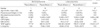
The characteristics of the enrolled children are presented in Table 1. Out of the 29 children with pneumonia, there were 16 children with bronchopneumonia (1 with parapneumonic effusion) and 13 children with lobar pneumonia (4 with parapneumonic effusion). Thoracentesis was performed in 4 patients with parapneumonic effusion except for one patient with bronchopneumonia who had only a scanty amount of fluid. All the pleural fluids were confirmed as exudates.
The mean age of the control group was higher compared to the pneumonia groups. However, age did not influence the serum VEGF concentrations. Neither our previous study (13) nor this study showed any correlations between serum VEGF levels and the subjects' age in control groups. The mean white blood cell counts in the patients with pneumonia were higher than those in the control subjects, but showed no differences among the pneumonia groups. The mean level of C-reactive protein was significantly increased in the patients having lobar pneumonia with effusion compared to the patients having lobar pneumonia without effusion and the patients having bronchopneumonia with effusion. The mean erythrocyte sedimentation rate was significantly increased in the patients having lobar pneumonia with effusion compared to the other groups, and the ESR was also significantly increased in the patients having lobar pneumonia without effusion compared to the patients having bronchopneumonia with effusion.
The urinary Streptococcus pneumoniae antigen test was positive in 5 of 13 patients with lobar pneumonia, and this test was negative in all the patients with bronchopneumonia. Among those five patients with positive tests, three patients had parapneumonic effusions. There were no culture-confirmed cases.
There were 8 patients with mycoplasma pneumonia. Four of them had bronchopneumonia and two patients had lobar pneumonia without effusion and two patients had lobar pneumonia with effusion. The serum levels of IL-6 showed no significant differences among the pneumonia groups.
Serum VEGF and IL-6 levels according to the radiologic types of pneumonia and the etiologies
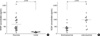
The mean concentration of serum VEGF was 669.42±85.28 pg/mL in the patients with pneumonia, and this was significantly increased compared to the control subjects (110.23±4.77 pg/mL, p<0.01). The mean level of serum VEGF in the patients with lobar pneumonia was 987.95±133.10 pg/mL, and this was significantly higher than that in the patients with bronchopneumonia (410.62±55.96 pg/mL, p<0.01) (Fig. 1).
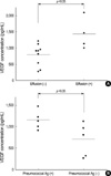
In the patients with lobar pneumonia, the serum VEGF levels of those with pleural effusion (1432.48±242.94 pg/mL) were also significantly higher than in those patients without pleural effusion (790.37±122.63 pg/mL, p=0.021). Likewise, in the patients with lobar pneumonia, the mean level of serum VEGF in those patients with a positive urine S. pneumoniae antigen test (1153.73±95.35 pg/mL) was significantly increased compared to those patients with negative results (702.06±172.15 pg/mL, p=0.047) (Fig. 2). For the patients with mycoplasma pneumonia, they did not show any significant differences in the serum VEGF and IL-6 levels compared to those patients without mycoplasma pneumonia (data not shown).
For the serum IL-6 levels, there were no significant differences between the patients with bronchopneumonia and lobar pneumonia, and between the patients with effusion and without effusion, and nor were there significant differences between the subjects who were positive or negative on the urinary S. pneumoniae antigen test (data not shown). The serum levels of VEGF in the patients with pneumonia showed positive correlation with their erythrocyte sedimentation rates (p=0.024), but there was no correlation with the serum IL-6 and CRP levels (Fig. 3). The white blood cell counts and the duration of fever showed no correlations with the serum VEGF levels (data not shown).
DISCUSSION
In this study, we have demonstrated that the serum VEGF concentrations were elevated in the children with pneumonia, and especially in those children who had lobar pneumonia and parapneumonic effusion. This may reflect that an acute lower respiratory tract infection can increase the production and release of VEGF. VEGF is known to be elevated in chronic lung diseases such as asthma and cystic fibrosis (7,8,14,15), whereas there are little data on its relationship with acute pneumonia (11), and furthermore, there is no data on CAP. In a study on acute eosinophilic pneumonia, the VEGF levels were measured in the bronchoalveolar lavage (BAL) fluids and its levels in the patients with acute eosinophilic pneumonia were higher than those VEGF levels of the control group, and the VEGF levels rapidly decreased to the control levels in parallel with the clinical improvement (11). Another clinical study showed that the serum VEGF levels were elevated in cystic fibrosis patients having acute exacerbations, and these levels were decreased with antibiotic therapy (14). These data support our results showing that the VEGF elevation is related to the airway inflammation associated with infection. On the contrary, in a study using lung autopsy material from septic patients, it was demonstrated that the pulmonary VEGF expression was decreased in the septic patients compared to that in the controls (16). Even though pneumonia, pulmonary edema and acute respiratory distress syndrome are the usual pulmonary manifestations during sepsis, the postmortem lung tissue seems to be different from the lung tissue seen in the acute infection states, and other factors such as mechanical ventilation, prolonged antibiotic use and a downhill clinical course to death will contribute to the confounding variables.
There have been no data that the serum VEGF level is related to the radiologic type of pneumonia. In this study, the serum levels of VEGF were higher in the patients with lobar pneumonia than in those patients with bronchopneumonia. It is not certain whether the VEGF elevation is related to the extent of inflammation or if it is related to the virulence of the microorganisms. In an in vitro study, Staphylococcus aureus stimulated VEGF release from the normal mesothelial cells in a dose-dependent and time-dependent pattern (17), and Streptococcus pneumoniae induced the dose- and time-dependent secretion of VEGF from human neutrophils (18). S. aureus and S. pneumoniae are two of the most common causes of severe, complicated pneumonia with parapneumonic effusion and empyema. There was no data on the association of the VEGF release with adenovirus infection, which can cause complicated pneumonia that mimics bacterial pneumonia (19).
In our study, the children with a positive urinary S. pneumoniae antigen test showed higher levels of serum VEGF than those children with negative results for lobar pneumonia. We found that all the cases with positive urinary pneumococcal antigen tests were lobar pneumonia with or without parapneumonic effusion; and no bronchopneumonia cases were positive on this test. Even though the urinary S. pneumoniae antigen test alone cannot be a tool for the definitive diagnosis of pneumococcal infection, it can be used as a presumptive diagnostic method, especially in case of invasive pneumococcal infection (whether it is bacteremia or lobar pneumonia). S. pneumoniae is isolated from the blood or pleural fluid in only 10-30% of pneumococcal pneumonia patients. The urinary S. pneumoniae antigen test yields a sensitivity of 77-92% and a specificity of 97-100% (20-23).
The level of VEGF has been shown to be consistently higher in the exudative pleural effusions than in the transudative pleural effusions (17,24-26). The empyema fluids also contain high levels of VEGF (24,27), which is up to five fold higher than the VEGF levels in the uncomplicated parapneumonic effusions (17). This study suggests that more complicated pneumonia which is usually caused by more virulent pathogens seems to be associated with a greater release of VEGF.
We did not compare the serum VEGF levels with those VEGF levels in the BAL fluid or the pleural effusion. The relative contribution of local VEGF production in pneumonia and also in the parapneumonic effusion is not well known. It was reported that local production is the main source of VEGF in the effusions rather than diffusion from the serum (6) and VEGF is likely to originate from multiple cellular sources such as the residential mesothelial cells, the circulating inflammatory cells, and the infiltrating malignant cells within the pleural space (28). There was the correlation of VEGF with the monocyte and macrophage concentrations (29). It is thought that during acute infection such as bacterial pneumonia, the blood supply increases as the inflammation progresses and the inflammatory cells infiltrate into the inflamed tissue. As a result of the local VEGF production by the recruited inflammatory cells, as well as the VEGF production from the pulmonary epithelial cells, the endothelial cells and the smooth muscle cells, the vascular permeability increased; this in turn causes pulmonary edema or effusion. In the systemic circulation, these inflammatory cells produce VEGF, and this causes the elevation of the serum VEGF levels as well.
In this study, serum VEGF levels increased significantly in the patients with pneumonia compared to the controls, especially in those patients with lobar pneumonia compared to those patients with bronchopneumonia. For the patients with lobar pneumonia, those patients with parapneumonic effusion showed higher levels of serum VEGF compared to the patients without effusion, and the children with a positive urinary S. pneumoniae antigen test showed higher concentrations of serum VEGF compared to those with negative results.
VEGF may be one of the key mediators that lead to more severe and complicated pneumonia; this can provide researchers with a new strategy for therapy in the future and so help in the prevention of severe and complicated pneumonia.

 XML Download