

 PDF
PDF ePub
ePub Citation
 Print
Print


INTRODUCTION
Benign paroxysmal positional vertigo (BPPV) is a common disorder wherein brief episodes of vertigo and nystagmus are produced by certain changes of head position (1-3). Dislodged otolithic debris may cause this disorder by attaching to the cupula and rendering it sensitive to head position (cupulolithiasis)(4), or by entering one of the semicircular canals and inducing endolymph flow by head motion (canalolithiasis) (5,6). The diagnosis of BPPV depends on characteristic history of positioning vertigo and on typical patterns of nystagmus induced by the positional changes including Hallpike maneuver or turning the head side to side in the supine position (head turning test).
BPPV most commonly involves the posterior semicircular canal. During Hallpike maneuver, the nystagmus of the posterior canal type of BPPV (PC-BPPV) is typically torsional upbeating with the torsional component beating toward the lowermost ear (1). The nystagmus in the horizontal canal type of BPPV (HC-BPPV) may beat toward the ground (geotropic) (-9) or away from the ground (apogeotropic) (10) while the head is turned to either side in the supine position. The geotropic type of HC-BPPV has been explained by canalolithiasis while the apogeotropic type has been ascribed to cupulolithiasis. Recently, torsional downbeat nystagmus during Hallpike maneuver has been invoked to explain BPPV from the anterior semicircular canal (AC-BPPV) (11-14).
As the canalith repositioning maneuvers (CRM) can result in rapid resolution of the symptoms and different maneuvers should be applied to each patient depending on the canal involved (15), delineation of the affected canal is very important in the diagnosis of BPPV. To characterize demographic features, patterns of canal involvement, and response to CRM in BPPV, we retrospectively analyzed the profiles of the patients, who had been diagnosed as having BPPV by trained neuro-otologists in seven Dizziness Clinics in Korea.
MATERIALS AND METHODS
Subjects
We reviewed the records of patients with a diagnosis of BPPV from seven Dizziness Clinics, and identified 1,692 patients who met the criteria of brief episodes of positional vertigo, and characteristic paroxysmal positional nystagmus confirmed by the neuro-otologists or recorded with oculography. Recruitment of the patients had been done from May 1997 to September 2003, with the recruitment periods varying from 13 to 76 months among the clinics.
Inclusion and exclusion criteria
Diagnosis of PC-BPPV was made by the torsional upbeating nystagmus with the torsional component toward the lowermost ear during Hallpike maneuver (1). HC-BPPV was subdivided into canalolithiatic (geotropic) and cupulolithiatic (apogeotropic) types according to the nystagmus direction induced by head turning while lying down (7-10). Paroxysmal torsional downbeating nystagmus with typical patterns of BPPV during Hallpike maneuver was classified into AC-BPPV (11-15). In cases of the nystagmus suggesting that two or more canals were involved (different patterns of the nystagmus depending on the provoking positional maneuvers), the mixed type of BPPV was diagnosed. Patients who reported a typical history suggestive of BPPV but did not exhibit positional nystagmus during the positioning maneuvers were excluded. In most patients with AC-BPPV and mixed type BPPV, when the repeated CRM did not result in resolution of the symptoms and signs, brain imaging was performed to rule out central pathologies. The patients with paroxysmal positional nystagmus accompanied by related CNS lesions were also excluded.
Treatment
For PC-BPPV, modified Epley procedure was advocated by most of the neuro-otologists. In canalolithiatic type of HC-BPPV, barbecue rotation maneuver was applied in most clinics. However, in some clinics and in a few patients with limitation in motion or without response even with repetitive rotation maneuvers, forced prolonged position was applied (16,17). In patients with cupulolithiatic type of HC-BPPV, most clinics performed head shaking maneuver first, which was followed by CRM if transition to canalolithiasis was noted. In a few clinics, vibratory stimuli on the mastoid process were also tried. In some patients with AC-BPPV, modified Epley maneuver was tried after Hallpike maneuver with an assumption that the otolithic debris were in the anterior canal of the ear toward the torsional component of the positioning nystagmus beat. In view of the frequent spontaneous resolution, responses to CRM were determined in only the patients who had the follow-up examination within 2 days after the initiation of CRM.
Analyses
Study of demographic and clinical characteristics was focused on the type of BPPV and response to CRM. Statistical analyses included chi-square test for dichotomous variables and ANOVA for continuous variables for comparison among the clinics. We used Pearson or Spearman correlation analysis to seek the correlation between the proportion of the involved canals and variables. A significance level of 0.05 was adopted.
RESULTS
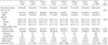
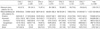
The demographic features were summarized in Tables 1, 2. BPPV was more common in female irrespective of the subtypes (female to male ratio of 2.1:1). The patients' mean age was 54.8±14.0 yr. There was no difference in the mean age between women and men, and among the subtypes. The mean time interval between the onset of the symptoms and the first visit to the clinics was 13.8±120.0 days. Eighty-nine percents of the patients visited the clinics within 14 days after the symptom onset.
The posterior canal was involved in 1,031 patients (60.6%, Table 1). HC-BPPV was found in 539 (31.9%) patients. Of HC-BPPV, 50.5% featured direction-changing apogeotropic nystagmus, while 49.5% showed geotropic nystagmus. AC-BPPV was identified only in 38 (2.2%), and mixed types in 84 (5.0%) patients. Among the mixed types, the combination of PC-BPPV and HC-BPPV was the most common (79.8%). There was significant difference in the distribution of involved canals among the clinics (p<0.05). We observed significant negative correlation between the proportion of HC-BPPV of each clinic and the mean time interval from the symptom onset to the first visit to the clinic (r=-0.841, p<0.05). In contrast, significant positive correlation was noted between the proportion of PC-BPPV and the mean time interval (r=0.845, p<0.05). However, no correlation was present between the mean age or sex proportion of the patients in each clinic and the proportion of each subtypes of BPPV.
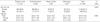
In 78.7% of the patients, CRM was performed (Table 3). Among the patients received CRM, 86.9% showed remission within two days. Geotropic type of HC-BPPV was most responsive (92.0%) while AC-BPPV was most refractory (76.2%).
DISCUSSION
Our study showed demographic features of BPPV similar to those reported previously in other countries; higher incidence in women and mean age of 54.8±14.0 yr (1,16,18). There was no difference in the age between women and men, and among the subtypes. Higher incidence in women was found in all the subtypes of BPPV.
As in the previous studies, the posterior canal was most commonly involved. However, the proportion of HC-BPPV was higher (31.9%) than previously reported. Most of the previous studies reported the proportion of HC-BPPV between 5 and 20% (19-22). Considering that the proportion of HC-BPPV was more than 25% in all the Clinics except for one, the higher proportion of HC-BPPV in our study cannot be ascribed to an incidental finding or diagnostic bias. The reason for this high proportion of HC-BPPV in our study remains to be elucidated. Due to the relatively unrestricted medical referral system in Korea, patients can visit the neurootologic clinics readily without delay. Actually, most of the patients in our study visited the Clinic within 14 days after the symptom onset. If the HC-BPPV has higher rate of spontaneous resolution compared with other types of BPPV (8,23), the delayed diagnosis would result in lower proportion of HC-BPPV, even though the exact interval from the symptom onset to the diagnosis was not available in most of the previous studies. This seems to be reasonable if one considers the spatial orientations of each semicircular canal. In upright head posture, the posterior canal hangs inferiorly and has its cupular barrier at its shorter, more dependent end. Any debris entering the canal essentially becomes trapped within it. In contrast, the horizontal canal slopes up and has its cupular barrier at the upper end. Therefore, free-floating debris in the horizontal canal would tend to drift back into the utricle more easily. This explanation is also supported by the highest proportion of HC-BPPV in the clinic (Clinic 1) which had the shortest time interval from the symptom onset to the clinic visit (Table 1). A recent study with a mean interval of 124 days from the symptom onset to the treatment, much longer than ours, reported the proportion of HC-BPPV at 8% (19). Another study in Japan also showed higher proportion of HC-BPPV (30%) as in Korea (24). However, due to the interval from the symptom onset to the diagnosis was not available in that study, we could not determine whether the similar distribution originated from the ethnic origin or time interval from the symptom onset to the diagnosis.
The diagnosis of AC-BPPV and mixed type of BPPV is challenging, and central pathologies should be ruled out appropriately (25,26). The characteristics of AC-BPPV have been described only recently. Previously, the proportion of AC-BPPV has been reported up to 12% (11). In our study, the mean proportion of AC-BPPV was 2.2% with the highest being 5.3%. Concurrent involvements of more than one canal had also been recognized, with the combination of PCand HC-BPPV being the most common (79.8%).
In view of the frequent spontaneous resolution of BPPV, we adopted a time window of 2 days to determine the efficacy of CRM, and demonstrated high efficacy of this maneuver, irrespective of the subtypes of BPPV. According to the previous studies, the Epley maneuver has been effective in 70 to 100% of patients suffering from this disorder (27-29). In our study, most neuro-otologists advocated modified Epley maneuver for the treatment of PC-BPPV, which showed resolution of the symptoms and signs in 87.8% of patients. Geotropic type of HC-BPPV also showed resolution in 92% of patients with the barbeque rotation maneuver or force prolonged posture. In spite of the debates on the efficacy of head shaking or mastoid vibration before and during CRM, those were applied to the patients with presumed cupulolithiatic type of BPPV in some clinics.
 XML Download