

 PDF
PDF ePub
ePub Citation
 Print
Print


INTRODUCTION
Corticotropin releasing factor (CRF) is a 41 amino acid peptide that is highly expressed throughout areas of the central nervous system (CNS). In the adult, CRF has been shown to be involved in the stress response mediated by the hypothalamo-pituitary-adrenal (HPA) axis, as well as being involved in some degenerative brain disorders (1-3). In adult mouse cerebellum, CRF has been localized to two major afferent systems, climbing fibers and mossy fibers, and it has been shown to function as a neuromodulator in adult cerebellar circuits (4-6).
Several observations suggest that CRF may have a trophic function in the developing cerebellum, as compared to its neuromodulatory role in the adult cerebellum. In the developing mouse cerebellum, CRF is initially detected at embryonic day 10 (E10) in widely dispersed profile, and is present throughout all pre- and postnatal stages of cerebellar development; however, its localization in phenotypic axon terminals (climbing and mossy fibers) begins to emerge at postnatal day 10 (P10) (7,8). Although CRF receptors (CRF-Rs) also are present on populations of neurons and glia at birth, a physiological effect of CRF on Purkinje cell firing rate is recorded at P12-P16 (8-10). Of particular note is a widespread distribution of CRF throughout cerebellum during embryonic and early postnatal development (8). The period between the initial appearance of CRF in cerebellum and its localization in cerebellar afferent fibers is critical in cerebellar development (7). In addition, proliferation or apoptosis of immature cerebellar neurons is found during this period.
CRF exerts its effects by binding to either the type 1 (CRF-R1) or the type 2 CRF receptor (CRF-R2). The high affinity receptor, CRF-R1, has one functional and several non-functional splice variants (11). Although CRF-R2 has three functional splice variants that are referred to as CRF-R2α, CRFR-2β, and CRF-R2γ, only CRF-R2α has a major distribution in the central nervous system (12). We will designate the functional splice variant of CRF-R1 and CRF-R2α as CRF-R1and CRF-R2, respectively, in this paper. Each receptor subtype has unique binding characteristics (13), and both CRF-R1and CRF-R2are differentially localized in mouse cerebellum during development (14-16). These results support that cerebellar neurons or astrocytes can be affected by ligands for CRF receptors during development. Our working hypothesis is that CRF or its family of peptides has a role during development that is distinct from that in mature brain, possibly acting as a trophic factor that influences neuronal survival and/or differentiation during cerebellar development. Previous reports have shown that CRF has a neuroprotective effect when neurons are exposed to a hypoxic or neurotoxic environment (17,18), while it has a mitogenic effect on immature astrocytes (19).
Recently, several members of CRF family of peptides, including urocortin (urocortin I), urocortin II and urocortin III, were identified (20-22). Urocortin II and III are selective ligands for CRF-R2 (20-22), whereas urocortin may bind to either CRF-R1or CRF-R2, but with a higher affinity for CRF-R2 The detailed cellular localization of urocortin and urocortin II in the developing and adult cerebella are yet to be determined; however, based on studies on the distribution of urocortin positive neurons (23), there are several nuclei that could be extrinsic sources of urocortin to the cerebellum. One possible influence that CRF family of peptides might have on developing neurons is to increase survival. We tested this possibility by culturing cerebellar neurons from E18 mice in the absence and in the presence of CRF, urocortin or urocortin II. The findings presented here suggest that CRF or urocortin might have a role in promoting the survival of GABAergic interneurons during early cerebellar development.
MATERIALS AND METHODS
Primary cerebellar neuron culture
Primary cell cultures of cerebella from E18 mice were established following the methods of Fischer (24) and Schilling et al. (25). Briefly, eighteen days after mating, pregnant C57BL/6 mice were anesthetized using an intraperitoneal injection of 2.5% Avertin (0.2 mL/10 g). The fetuses were removed, decapitated, and placed in phosphate-buffered saline (PBS). The cerebella were dissected from the brainstem and the meninges were removed. The PBS then was replaced with Basal Medium Eagles solution (BME Complete Medium) with transferrin (100 µg/mL), aprotinin (1 µg/mL), selenite (30 nM), triiodothyronine (0.2 ng/mL), insulin (10 µg/mL), NaHCO3 (26.18 mM), glucose (0.25%), pyruvate (1 mM), glutamate (2 mM), and bovine serum albumin (BSA) (1.010 mg/mL) plus 5% heat-inactivated fetal bovine serum. The cerebella then were triturated 15 times before the cells were passed through a 40 µm cell strainer (BD Biosciences, Bedford, MA, U.S.A.). After centrifugation at 150×g for 5 min, the pellet was resuspended in BME Complete Medium with 5% heat-inactivated fetal bovine serum. Cells then were counted using a hemocytometer (Bright-line; Hausser Scientific, Horsham, PA, U.S.A.) and plated at a density of 100 ×103cells/cm2 into a four-chamber (1.8 cm2) culture slide (Lab-Tek; Nalge Nunc International, Rochester, NY, U.S.A.) that was pre-treated with poly-D-lysine (10 mg/100 mL). The cultures were incubated in 10% CO2 at 37℃. One day after plating (at 1 day in vitro; 1 DIV), the cells were fed with serum-free BME Complete Medium containing the various agents as delineated below. Every 3 days, half the volume of medium was removed and replaced with fresh complete medium containing the same appropriate agents. All procedures involving animals have been approved by Sungkyunkwan University School of Medicine Institutional Animal Care and Use Committee and conform to NIH guidelines.
Each culture was treated for either 1 day, 5 days or 9 days with one of the following conditions: a) BME complete medium only (control), b) BME complete medium with 1 µM CRF, c) BME complete medium with 1 µM urocortin, d) BME complete medium with 1 µM urocortin II, e) BME complete medium with 1 µM astressin or f) BME complete medium with 1 µM astressin in the presence of either 1 µM CRF, 1 µM urocortin or 1 µM urocortin II. At 1, 5 or 9 days of treatment (DT), cultures were fixed with 4% paraformaldehyde solution for immunocytochemistry. A minimum of three independent experiments per each condition were performed. We have tried several cell plating densities for cultures and found that 100×103cells/cm2cell plating density is suitable for demonstrating the effect of CRF and its family of peptides. The serum-free culture medium (BME complete) used in the study has been shown previously to suppress cell proliferation, to support the survival of neuronal cells, to have less than 5% of astrocytes in relation to total number of cells, and to eliminate epithelioid cells such as fibroblasts (24).
Immunocytochemistry
Cells in culture were identified using double-label immunofluorescence technique. Briefly, cultures were fixed with 4% paraformaldehyde, 0.1 M phosphate buffer (pH 7.1), 50 mM sucrose, and 0.4 mM CaCl2 for 30 min, and then rinsed with PBS+(PBS containing 0.1% saponin, 0.02% sodium azide, and 1 mg/mL BSA). Cells were incubated in primary antibodies at 4℃ overnight, rinsed with PBS+, and incubated in species-specific secondary antibodies for 2 hrs at room temperature. After rinsing with PBS+, slides were coverslipped with Immu-Mount and sealed with clear fingernail polish to avoid dehydration.
Image acquisition and analysis
Using a Zeiss Axiophot 2 fluorescence microscope (Zeiss, Germany) and Axiovision 3.0 Image Acquisition System (Zeiss, Germany), the acquisition time and gain were standardized to obtain the optimal images. For each slide, a series of consecutive digital images following a horizontal line (180 µm2/sample, spanning the width of each chamber) were acquired to ensure that a uniform area was analyzed and an adequate sample size for quantification was obtained. The number of GABA-positive neurons was counted for monitoring the changes in neuronal survival and the cell counts were expressed as means±standard deviations (a minimum of 400 cells per condition). Significance was determined using Student's t-test, two-tailed.
Chemicals and antibodies
The following chemicals were obtained from Sigma: aprotinin, BSA, glucose, glutamate, insulin, lysine, pyruvate, saponin, selenite, sodium bicarbonate, sodium chloride, sodium phosphate, sucrose, triiodothyronine, and transferrin.
The following chemicals were obtained from various sources as indicated: Basal Medium Eagles (BME) (Invitrogen-Gibco, Carlsbad, CA), human/rat CRF (Bachem, San Carlos, CA), urocortin (Bachem, San Carlos, CA), urocortin II (Bachem, San Carlos, CA), astressin (Bachem, San Carlos, CA), fetal bovine serum (Hyclone, Logan, UT), Immu-Mount (Thermo Shandon, San Jose, CA), sodium azide (EM Sciences, Hatfield, PA), and paraformaldehyde (EM sciences, Hatfield, PA).
Primary antibodies were used as follows: rabbit anti-GABA IgG (1:1,000) (Sigma), goat anti-CRF-R1(1:500) (Santa Cruz Biotechnology, Santa Cruz, CA) and goat anti-CRF-R2(1:500) (Santa Cruz Biotechnology, Santa Cruz, CA). Fluorescen secondary antibodies were used as follows: Alexa 488-labeled donkey anti-rabbit IgG (Molecular Probes, Eugene, OR) or Alexa 546-labeled donkey anti-goat IgG (Molecular Probes, Eugene, OR) was used in combination with anti-GABA or anti-CRF receptor antibodies, respectively. All secondary antibodies were used at 1:1,000 dilutions.
RESULTS
General appearance of cultures
To determine if CRF or its family of peptides enhances the survival of GABAergic neurons in growth-promoting culture medium, we used primary cell cultures using cerebellar cells from E18 mice because neurons that are known to be GABAergic have been born by this age and these cultures have been reported to contain a variety of cerebellar GABAergic interneurons including basket cells, stellate cells, and/or Golgi cells, which are labeled with anti-GABA antibody. Immunocytochemistry indicated that our cultures contained numerous GABAergic interneurons and only a few scattered Purkinje neurons, identified by calbindin immunocytochemistry (data not shown). To determine if the control culture condition (BME complete) affects the survival of GABAergic interneurons even without treatment with CRF family of peptides, cell counts were made for GABAergic interneurons at each culture age. As for plating density of cells in culture, our preliminary studies indicated that higher plating densities than 100×103 cells/cm2 masked the effect of CRF, possibly due to endogenous factors that promote cell survival; increasing the plating density to 150×103 cells/cm2 partially reduced the observed effects, while little effect was observed when the plating density was 200×103 cells/cm2 or higher. Based on the previous data, this study was conducted using a plating density of 100×103 cells/cm2.
The survival and maturation of GABAergic interneurons in control cultures
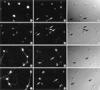
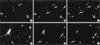
Images of the control cultures at each culture age are shown in Fig. 1. The immunocytochemical labeling for GABA shows that our cultures have a significant number of GABAergic interneurons at each culture age, thus, these cultures can serve as a good model for studying the influence of possible trophic factors on cerebellar GABAergic interneurons. At 1 DIV (Fig. 1A), most GABAergic neurons in culture are present isolated from each other and show immature morphology. Most GABAergic neurons do not yet show any prominent neurite growth at this culture age, and only a few neurons have short neurites growing from their somata, which appear as filopodia or lamellopodia. At 2 DIV, the number of GABA-immunopositive neurons in control cultures decreases to 60% of that at 1 DIV (Fig. 2). Some GABAergic neurons show more differentiated morphology with one or two neurites around their somata, and neuronal cell bodies are present adjacent to each other (Fig. 1D, 3A). At 6 DIV, the number of surviving GABAergic interneurons further decreases compared to 2 DIV (Fig. 2), but most neurons show more mature morphology with multiple long neurites around their somata (Fig. 1G). In addition, there is extensive growth of neurites on the substrate between neurons at 6 DIV; the neurites come in very close proximity to, or make contact with, each other at 6 DIV. At 10 DIV, the number of GABA-immunopositive neurons does not further decrease compared to 6 DIV (Fig. 2). The GABAergic interneurons in cultures show fully mature morphology with highly branched long neurites, which form an extensive network between multiple neurons (Fig. 1J, 3D). Based on these observations, it is evident that these culturing conditions will permit growth of healthy cerebellar GABAergic interneurons.
CRF receptor expression in cultured cerebellar GABAergic interneurons
For a neuron to be responsive to some kind of ligands, it should have specific receptors on their surface. To verify the presence of CRF receptors on neurons under these culturing conditions, cerebellar cultures were fixed at 1, 2, 6, and 10 DIV and labeled with antibodies to CRF-R1or CRF-R2. The representative images, from the same fields as shown in Fig. 1A, 1D, 1G, 1J, 3A and 3D are presented in Fig. 1B and 1C, 1E and 1F, 1H and 1I, 1K and 1L, 3B and 3C, and 3E and 3F, respectively. At 1 or 2 DIV in control culture, the majority of GABA-immunopositive neurons are also positive for CRF-R1(Fig. 1B; 1 DIV, Fig. 1E; 2DIV) or CRF-R2(Fig. 3B; 2DIV), indicating that they have the ability to respond to CRF or its family of peptides. There exist some CRF-R1or CRF-R2immunopositive cells that do not show labeling for GABA (Fig. 1B, 1E, 1H, 1K, 3B and 3E, white arrows; Fig. 1C, 1F, 1I, 1L, 3C and 3F, black arrows); these CRF-R1or CRF-R2positive, but not GABA immunopositive cells are supposed to be another kind of cerebellar neurons, the granule cells, because those cells are not labeled for calbindin, a marker for Purkinje cells, nor are they labeled for glial fibrillary acid protein (GFAP), a marker for astrocytes (data not shown). Further study is yet needed to identify these cells that are not labeled for GABA, calbindin or GFAP. Most GABAergic neurons appear to be also positive for CRF-R1and CRF-R2under all experimental conditions from 1 DIV to 10 DIV, indicating that the treatments with CRF family of peptides do not alter the expression of CRF receptors in these cultures.
Survival of GABAergic interneurons in treatment with CRF family of peptides
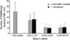
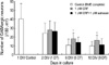
Based on our hypothesis, one putative role for CRF and its family of peptides is that they act to influence cell survival during cerebellar development. Therefore, cultures were treated for one, five or nine days with 1 µM CRF (Fig. 4), 1 µM astressin (Fig. 2), 1 µM urocortin (Fig. 5) or 1 µM urocortin II (Fig. 5). While the overall health of the cultures in each experimental condition appears to be fine, the number of surviving GABAergic neurons is altered in specific treatment conditions as the duration of treatment increases (at 5 DT and 9 DT).
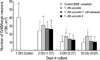
Treatment with 1 µM CRF for 1 day (1 DT, at 2 DIV) does not show any change in the survival of GABAergic neurons in culture compared to control culture (BME complete) (Fig. 4). However, treatment with CRF begins to show a significant increase in the survival of GABAergic neurons at 5 DT (6 DIV) when compared to the control culture (BME complete), and the increased survival of GABAergic neurons is maintained through 9 DT (10 DIV). At 5 DT with 1 µM CRF, the number of GABAergic interneurons is 66% higher than control level, and this value appears to be statistically significantly higher, suggesting a role for CRF that might promote the survival of cerebellar GABAergic neurons in cultures. At 9 DT with 1 µM CRF, the average number of surviving GABAergic neurons is 63% higher than control level, and this value also appears to be significantly higher in statistics, suggesting that the survival promoting effect of CRF in our cultures is maintained through this culture age. To confirm that the survival promoting effect at 5 DT and 9 DT is actually mediated by 1 µM CRF treatment, we treated the cultures with 1 µM astressin, the specific antagonist to both subtypes of CRF receptor, in the presence of 1 µM CRF. As a result, 1 µM astressin treatment abolished the survival promoting effect of CRF at both 5 DT and 9 DT (Fig. 4); statistical analysis for the effect of 1 µM astressin treatment in the presence of 1 µM CRF showed that there was no statistically significant difference in GABAergic cell numbers between treated culture and control culture at both 5 DT and 9 DT. Higher concentration of antagonist, 10 µM astressin, also has been employed to compare the antagonistic effect between various concentrations of astressin in the presence of 1 µM CRF, and as a result, 10 µM astressin also appeared to be effective to abolish the survival promoting effect of CRF (data not shown). It is possible that astressin also can affect the survival of cerebellar GABAergic interneurons in cultures, although it is not a physiological agent that is naturally present in vivo. Thus, to determine any possible effect of astressin on the survival of GABAergic neurons in vitro, we treated cultures with 1 µM astressin alone in BME complete media. The presence of astressin at 1 µM in cultures did not show any significant change on the survival of GABAergic neurons, compared to control (Fig. 2); therefore, we can suggest that in our cultures, astressin abolished the survival promoting effect of CRF by antagonizing CRF receptors.
In addition to CRF, we also treated cultures with other members of CRF family of peptides, urocortin and urocortin II. Cultures treated with 1 µM urocortin resulted in a significant increase in the survival of GABAergic interneurons at 5 DT and 9 DT compared to control (Fig. 5), whereas treatment treatment with 1 µM urocortin II did not result in any significant change in cell survival at all culture ages (Fig. 5). To confirm that the survival promoting effect of urocortin was mediated through CRF receptors, we treated cultures with 1 µM astressin in the presence of urocortin. As a result, 1 µM astressin treatment abolished the survival promoting effect of 1 µM urocortin at both 5 DT and 9 DT (Fig. 5); statistical analysis of the effect of 1 µM astressin treatment in the presence of 1 µM urocortin showed that there was no statistically significant difference between treated culture and control culture, which suggests that the survival promoting effect was made by treatment with 1 µM urocortin through CRF receptors.
DISCUSSION
E18 mice cerebella, which are used in this study for culturing neurons, contain populations of cerebellar neurons including post-mitotic Purkinje cells; GABAergic interneurons including basket, stellate, and Golgi cells; nuclear neurons; and immature granule cells; as well as populations of primitive astrocytes and Bergmann glia (26). Because neuronal survival following the dissociation procedure is the greatest for cell populations that have not yet developed extensive neurites and are still undergoing mitosis and/or migration, the neurons that are most likely to survive in our cultures include the GABAergic interneurons, i.e., basket cells, stellate cells, and Golgi cells. While most of these GABAergic interneurons are born between E12 and E15 in the ventricular zone, data show that these neurons continue to divide as they migrate through cerebellar white matter and cortex (27); therefore, these neurons still remain in an immature state at E18 when they are used for culture, and as a result, many cells survive in culture. We confirmed that most neurons that survived in our culture system following dissociation procedure were GABAergic interneurons by doing immunocytochemistry for GABA.
In a previous study in which cerebellar cells were plated at a high density and grown in media containing serum, astrocytes without neurons were present in culture, and CRF treatment showed an increased number of astrocytes compared to cultures grown without CRF (19). In our cultures, we eliminated glial cells by plating cells at a low density, as well as using culture media that contained no serum, but contained a supplement that allowed neuronal survival. In addition, the low plating density used in our cultures has an advantage in minimizing the influence of endogenous factors produced by other cells in culture, while it still allows GABAergic interneurons to survive.
The decrease of GABAergic interneurons in control cultures is the greatest during the first day following removal of serum from the culture media. There should be many cells that were damaged during dissociation procedure, and as a result, those cells would die rapidly within a few days following cell plating. At 6 DIV, the number of GABAergic interneurons further decreases in control culture. The decrease of GABAergic neurons during this period may still represent death of cells that were damaged during dissociation procedure, or partially represent death of cells that can not obtain enough endogenous factors released from other cells, including glia, due to the low plating density used in our culture. However, the number of GABAergic neurons does not further decrease after 6 DIV, and almost the same number of GABAergic neurons survive at 10 DIV. This finding may represent that cells that were damaged during dissociation procedure or poorly supplied with endogenous factors have been eliminated from culture by 6 DIV. Although only 25% of GABAergic neurons that were originally plated on the substrate survive at and after 6 DIV, our data show that enough number of GABAergic neurons needed for experiment finally survive in culture, so that this culture system is a good in vitro model for studying the trophic effect of CRF family of peptides on the survival of cerebellar GABAergic neurons.
Both subtypes of CRF receptors are expressed in the majority of GABAergic neurons in control cultures from 1 DIV through 10 DIV, and no obvious change is found in the intensity of immunolabeling between culture ages. Previous reports showed that the expression of CRF receptor was regulated reciprocally depending on the level of CRF that were present in local areas of brain (28). However, in our cultures, treatment with CRF family of peptides at 1 µM concentration does not induce any remarkable change in the expression level of CRF receptors on GABAergic neurons. Although the mechanism regulating the expression of CRF receptor in cultured neurons as well as in cerebellum during early period of development is not yet clear, the known mechanism by which CRF regulates the expression of CRF receptors in a reciprocal manner may not work in culture system.
Based on our data, 55% of GABAergic neurons have died in control culture during 4 days from 2 DIV to 6 DIV, but in the presence of 1 µM CRF, only 26% of neurons have died during the same period, which represents that 1 µM CRF treatment reduces neuronal death in culture. Considering this finding together with the possible causes for neuronal death during the first several days in culture, we can suggest that CRF may be effective to promote the survival of neurons that were not severely damaged or poorly supplied with endogenous factors. At 10 DIV, treatment with 1 µM CRF increases the neuronal survival by 63% when compared to control. Based on our data, the survival promoting effect induced by 1 µM CRF appears to be almost the same at both 6 DIV and 10 DIV. Considering that the average numbers of surviving GABAergic neurons are almost the same at both culture ages in control cultures, we can conclude that 1 µM CRF treatment does not induce proliferation of immature GABAergic neurons in cultures, but it shows a protective effect reducing cell loss that occurs following dissociation procedure or in a poor nutritional condition.
We treated cultures with other members of CRF family of peptides, urocortin and urocortin II, to investigate which subtype of CRF receptor is involved in the possible protective mechanism induced by CRF family of peptides, as well as to determine the effect of each peptide on cultured GABAergic neurons. Based on the ligand specificities for CRF-R1and CRF-R2, we can compare the role for each subtype of CRF receptor in mediating the effect of CRF family of peptides on cultured neurons. Based on our data, CRF-R1 may have a role in the mechanism that regulates the survival of cultured GABAergic neurons in the presence of CRF or urocortin, while CRF-R2may not have a role in the mechanism. Although our data show that urocortin is effective in promoting the neuronal survival in cultures, the detailed information about the spatial and temporal expression of urocortin and urocortin II during cerebellar development, as well as in adult cerebellum, is not yet clear; therefore, further study is needed to clarify the role for urocortins in cerebellum during development and in adult.
Our data indicate that treatment with 1 µM astressin abolishes the survival promoting effect of CRF or urocortin at both 6 DIV and 10 DIV. In addition, astressin does not have any trophic effect, nor does it have toxic effect on cultured GABAergic neurons. Considering the receptor specificities for CRF family of peptides as well as the role for astressin, we can suggest that the survival promoting effect of CRF and urocortin on cultured GABAergic neurons may be mediated by the type 1 CRF receptor.
Although the mechanism by which CRF or urocortin increases the survival of cultured GABAergic neurons is not yet known, one possible mechanism has been suggested recently by Radulovic et al. (29). They found that a cytoprotective effect of CRF in Y79 human retinoblastoma cells is exerted by suppressing pro-apoptotic pathways, probably at a site upstream from procaspase-3. Since GABAergic neurons in our cultures have been shown to express CRF-R1extensively, it is not inconceivable that the survival promoting effect of CRF or urocortin reported in this study might employ a similar mechanism. Additional study is needed to confirm such a hypothesis. The findings presented in this study suggest a possibility that CRF or urocortin is capable of influencing cerebellar development by increasing neuronal survival in GABAergic neurons when cells are apoptotic or poorly supplied with endogenous factors needed for survival or differentiation.
 XML Download