

 PDF
PDF ePub
ePub Citation
 Print
Print


INTRODUCTION
Perinatal asphyxia is an important cause of neonatal mortality and permanent neurologic sequelae such as cerebral palsy, mental retardation, learning disability, and epilepsy in survivors (1-3). As most asphyxial events occur before birth, it is practically impossible to institute some neuroprotective treatments during the primary hypoxic-ischemic insult. However, it has become clear that although cerebral hypoxia-ischemia may cause immediate cell death during the insult, it can also lead to delayed cerebral injury hours, days or even weeks later (4-6). Thus the brain damage ongoing during reoxygenation- reperfusion could give us a therapeutic window during which we may be able to intervene before delayed irreversible cell death occurs, and thus to inhibit or reverse permanent neonatal brain damage (3,5,7-9).
There are two major forms of cell death that can be distinguished during hypoxic-ischemic brain injury (10-13). Necrosis is a rapid passive form of cell death characterized by prominent acute cell body swelling with subsequent cell lysis. Apoptosis is a delayed active form of cell death characterized by compaction of the cell body and internucleosomal DNA cleavage, and requiring new protein synthesis (14). In an adult rodent model of middle cerebral artery occlusion (MCAO) with cycloheximide treatment, the therapeutic window, of up to 6 hr, existed only in the apoptotic cell death following mild ischemia, but not in the acute necrosis following severe ischemia (15). In the developing brain, apoptosis has been found to play an important role in mediating neurodegeneration in pathological conditions (4,5,9,12,16). Furthermore, newborn brain infarction developed and progressed in a much delayed fashion (17,18); apoptotic cells were still observed at the peri-infarct area even weeks after the hypoxic-ischemic insult (18). Therefore, these findings raise the intriguing possibility that a delayed anti-apoptotic therapeutic intervention might prove to be valuable in reducing neonatal hypoxic-ischemic brain injury.
In our previous study (18), we demonstrated that apoptosis plays an important role in the development of delayed brain injury, and cycloheximide even when given after hypoxia-ischemia (HI) inhibits apoptosis, and reduces the ensuing cerebral infarction in a newborn rat pup model. These results suggest that anti-apoptotic treatments may have a useful role as neural rescue therapies for newborn infants (5). However, enrollment of newborns for clinical trials can take several hours for a mother to recover from a delivery and to give informed consent, and it is practically impossible to start any therapy immediately after birth. As there is a danger that delay in applying potentially brain-saving treatments beyond the therapeutic window might abolish or drastically reduce its therapeutic effectiveness (3,7,8,19), better understanding of the therapeutic window must be obtained before translation of experimental results to clinical trials. The present study was thus performed to determine the therapeutic window for cycloheximide treatment on neonatal hypoxic-ischemic brain injury.
MATERIALS AND METHODS
Hypoxia-ischemia
The experimental protocols described herein were reviewed and approved by the animal care and use committee of the Samsung Biomedical Research Institute, Seoul Korea. This study also followed the institutional and National Institute of Health guidelines for laboratory animal care.
Unilateral carotid artery ligation was performed in 7-day old Sprague Dawley rat pups (Daihan Biolink Co., Seoul, Korea) under methoxyflurane anesthesia. The neck was incised in the midline, and the right common carotid artery was permanently ligated with 4-0 silk. Total time of surgery in each animal never exceeded 5 min. Following surgery, rats were returned to their mother for recovery and feeding for 2 hr. The pups were then exposed to a 100 min-period of hypoxia (8% O2, 92% N2) by placing them in an airtight chamber partially submerged in a temperature controlled water bath to maintain the ambient temperature inside the chamber at a constant 36℃. In the HI with cycloheximide treatment group, the rat pups received an intraperitoneal injection of cycloheximide at a dose of 0.6 mg/kg (20) at 0, 6, 12 or 24 hr of recovery, and an equal volume of normal saline was given to a HI control group. Then, the rat pups were returned to their dam until sacrifice; the whole brain tissue was obtained under deep pentobarbital anesthesia (60 mg/kg, intraperitoneal) for flow cytometry and triphenyl tetrazolium chloride (TTC) at 48 and 72 hr after HI, respectively. Eight animals were used in each subgroup of analyses.
Flow cytometry
To evaluate the extent of apoptotic and necrotic cells, the mid-portion of ipsilateral cerebral cortex was dissociated into a single cell, and flow cytometry was done with a combination of PI (Sigma, St. Louis, MO, U.S.A.) and annexin V fluorescein isothiocyanate (FITC) (Pharmingen, San Diego, CA, U.S.A.) at 48 hr after hypoxia-ischemia. The flow analysis was performed by a PAS (Particle analyzing system, Partec, Munster, Germany) equipped with an argon ion laser tuned at a 488 nm wavelength. The green FITC-annexin V fluorescence was measured at 530±15 nm, and the red PI fluorescence was measured at 600 nm (18,21).
Morphometric analysis of infarct volume
Infarct volumes were measured by morphometric analysis of infarct areas that were defined by TTC (Sigma, St. Louis, MO, U.S.A.) at 72 hr after HI (15). This method provides an overall measure of cell injury presented by depleted NADPH and hence the inability to reduce TTC to its colored form. After intracardiac perfusion with 20 mL of 0.9% NaCl, brains were cooled in ice-cold saline for 5 min, and then sliced into 1 mm thick coronal sections using rat brain matrices (Harvard Bioscience, South Natick, MA, U.S.A.). The brain slices were incubated in phosphate-buffered saline (pH 7.4) containing 2% TTC at 37℃ for 20 min, and then stored in 10% neutral-buffered formalin. A cross-sectional area of the TTC-stained region for each brain slice was determined using an Optimas 6.51 Image Analysis System (Media Cybernetics Inc., Silver Spring, MD, U.S.A.). After integration of these stained areas, the indirect method of Swanson was used to determine infarct volume (subtraction of residual right hemisphere cortical volume from the cortical volume of the intact left hemisphere) (22).
RESULTS
Flow cytometry
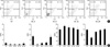
Representative analysis and regional percentage of an annexin V versus PI dot plot of ipsilateral cerebral cortex in HI control and HI with cycloheximide-treated groups (0, 6, 12, 24 hr after HI, respectively) at 48 hr after HI are presented in Fig. 1. When cycloheximide was given immediately after hypoxia-ischemia (0 hr), the percentage of damaged, necrotic and apoptotic cells in the respective area of Q1 (annexin V−/PI+), Q2 (annexin V+/PI+) and Q4 (annexin V+/PI−) decreased, and the live cells in Q3 (annexin V−/PI−) increased significantly compared to the HI control group. When cycloheximide was administered 6 hr after HI, the damaged and necrotic cells in Q1 and Q2 were significantly decreased, and the decrease of apoptotic cells in Q4 did not reach a statistical significance compared to the HI control group. With cycloheximide treatment at 12 hr after HI, only the necrotic cells in Q2 were significantly reduced compared to the HI control group, and no protective effect was seen if administration was delayed until 24 hr after HI.
Morphometric analysis of infarct volume
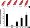
Infarct volume, as measured by morphometric analysis of infarct areas with TTC, was significantly reduced by 92% and 61% when cycloheximide was given 0 or 6 hr after HI respectively, but showed an insignificant trend in infarct reduction if cycloheximide was administered 12 hr after HI compared to the HI control group, and no protective effect was observed when administration was delayed until 24 hr after HI (Fig. 2).
In both flow cytometric analyses and infarct volume measurements, there was a trend of better neuroprotection with earlier administration of cycloheximide after HI.
DISCUSSION
In the present study, cycloheximide significantly attenuated the brain injury due to HI when given up to 6 hr after HI in a newborn rat pup model. These findings support the assumption that a therapeutic window exists in the early hours following a primary hypoxic-ischemic insult when intervention can attenuate irreversible brain damage in newborn infants with perinatal asphyxia (3,5,7-9). As the delay in administering a drug in humans beyond the therapeutic window might obscure or nullify its therapeutic effectiveness, time is a critical element for successful clinical trials (3,7,8,19). Furthermore, a trend of better neuroprotection with the earlier administration of cycloheximide after HI was observed in the present study. These findings suggest that an early selection of patients and administration of the treatment as soon as possible is of utmost importance for maximal neuroprotection in clinical trials (3,8).
In an adult rat model of MCAO (15), cycloheximide reduced infarct volume when given up to 6 hr after 30 min of mild transient ischemia. However, if the duration of ischemic insult was increased to 90 min, the therapeutic window for delayed cycloheximide was only 30 min, and even pretreatment of cycloheximide was ineffective in permanent MCAO. These findings further support a therapeutic window for cycloheximide treatment exists only in the apoptotic delayed form of cell death, but not for acute necrosis. The same therapeutic window of up to 6 hr with cycloheximide treatment observed both in the newborn rat pup model of the present study and in the adult model of MCAO (15), also suggests that the mode of neuronal death, apoptosis or necrosis, rather than the maturity of the brain itself are the important factors for this therapeutic window.
Further study will be necessary to elucidate the mechanisms underlying the cycloheximide-induced neuroprotective effects observed here. The reason why inhibition of protein synthesis should block apoptosis has not been completely understood, but presumably some critical protein must be synthesized de novo before apoptosis can proceed (14). However, actions of cycloheximide relevant to apoptosis other than the inhibition of death protein synthesis have also been recognized. Furukawa et al. (23) showed that cycloheximide treatment at low levels insufficient to fully inhibit protein synthesis enhances Bcl-2 expression. Cycloheximide can also attenuate free radical stress by inhibiting prostaglandin synthesis (24), and by increasing the availability of cysteine for glutathione synthesis (25).
In our previous (18) and present studies, significant reduction of both apoptotic and necrotic cells, measured by flow cytometry and brain infarction, was observed with cycloheximide treatment. As a working hypothesis, we think it is likely that inhibition of apoptosis with cycloheximide might also attenuate the secondary necrotic cells representing the secondary degradation of apoptotic cells (12,26), and the ensuing tissue necrosis (cerebral infarction). Administration of cycloheximide within the therapeutic window could attenuate activation of the neurotoxic, probably apoptotic, cascade ultimately leading to delayed cell death and irreversible brain damage hours, days or months later (3,5,7-9,15). Apoptotic neuronal death is a relatively slow and multi-step process, and the entire process, from the initial trigger to the destruction of the cell, can take hours or even days (4-6,27). However, our data demonstrating a therapeutic window less than 6 hr after HI to inhibit or reverse apoptosis with cycloheximide suggests that although the whole apoptotic process may be gradual and delayed, the fate of cells would be determined early after the insult, and once the apoptotic events proceed beyond the critical control point, the cell may become irreversibly committed to death (27,28).
In conclusion, cycloheximide treatment was effective in attenuating brain injury within a 6 hr therapeutic window after HI in a newborn rat pup model. Our data support the possibility that protein synthesis inhibitors, as well as other anti-apoptotic strategies, may have therapeutic utility in hypoxic-ischemic events of the developing newborn brain even when treatment is delayed for up to 6 hr after the primary asphyxial insult.
 XML Download