

 PDF
PDF ePub
ePub Citation
 Print
Print


INTRODUCTION
Among the important functions of B cells are antigen recognition, antigen presentation, antibody production, and immune regulation. The surface markers of B cells include immunoglobulin (Ig) M and IgD, which bind antigen, as well as CD19, CD20 and CD21 (1). Recent results suggest that the strength of the signal generated through the antigen binding receptor determines the fate of B cells, that is, whether they exist as immunoglobulin-secreting B-1 cells or as conventional B-2 cells (2,3).
The neck has complicated anatomic structures, with lymph node influx from the neck itself, the face, the oral cavity, the upper extremities, the chest, and the abdomen via the thoracic duct. The neck contains about 30% of the lymphoid tissue and lymph nodes in the entire body. Consequently, these lymphoid structures have a major role as a primary defense barrier against bacteria and other foreign pathogens and against the spread of malignant tumors. Although the pathology of lymph nodes has been extensively investigated, there have been few studies of normal lymph nodes (4,5). In addition, little is known about B cells of cervical lymph nodes, perhaps because these cells are considered identical to all other lymphoid B cells and because of the ethical issues involved in obtaining sufficient cervical lymph nodes of normal individuals. Cervical lymph nodes, however, are exposed to continual antigenic stimulation from the naso- and/or oro-pharynx. In the present study, we therefore compared the characteristics of normal murine cervical lymph node B cells with those of B cells of spleen and peritoneal fluid, which consist of B-2 and B-1 cells (6-8).
MATERIALS AND METHODS
Animals
Male BALB/cByJ mice 8-14 week of age were obtained and housed at least 1 week before experimentation. Mice were cared for and handled at all times in accordance with institutional animal care committee.
B cell purification and cell count
B cells were obtained from cervical lymph nodes, spleen and peritoneal fluid. Cell suspensions were depleted of T cells by treatment with anti-Thy-1.2 antibody plus rabbit complement. They were then plated on plastic dishes to deplete macrophage. Red blood cells and nonviable cells were removed by sedimentation over Lympholyte-M (Cedarlane, Ontario, Canada). The resulting B cells were cultured in Roswell Park Memorial Institute medium 1640 (BioWhittaker, Walkersville, MD, U.S.A.) supplemented with 5% heat-inactivated fetal bovine serum (Sigma-Aldrich, St. Louis, MO, U.S.A.), 10 mM Hepes (pH7.2) (Calbiochem-Novabiochem, San Diego, CA, U.S.A.), 50 µM 2-ME (Sigma-Aldrich), 2 mM L-glutamine (Invitrogen Co., Japan), 100 U/mL penicillin, and 100 µg/mL streptomycin (Invitrogen Co., Japan).
B cells then were divided into four groups; First, 0.5×10⁶ cells for their expression of B cell surface glycoproteins. Second, 2.0×10⁶ cells for cell survival rate. Third, 2.0×10⁶ cells for the in vitro ELISA assay to measure the amounts of immunoglobulin and B cell differentiation. Fourth, 1.0×10⁶ cells for cell proliferation assay to evaluate the entering of S phase.
Flow cytometry: cell surface staining
Purified B cells or lymphocytes from the cervical lymph node, spleen and peritoneal fluid were assessed by immunofluoresent staining and flow cytometric analysis (Becton Dickinson Corp., Sunnyvale, CA, U.S.A.) to determined the characteristic features of cells. For staining of B cells, B cells were incubated with fluorescein isothiocyanate (FITC)-conjugated monoclonal anti-CD220, phycoerythrin (PE)-conjugated monoclonal antibodies which anti-CD5, anti-CD23, anti-CD43, anti-CD80 (B7.1), anti-CD86 (B7.2), anti-IgM, or anti-CD 138 (Syndecan-1) (BD PharMinogen, San Diego, CA, U.S.A.).
Survival rate
Cell viability was determined by trypan blue exclusion (Sigma-Aldrich) and counting cells with a hemocytometer on days 2 and 5.
Enzyme-linked immunosorbent assay (ELISA)
Total secreted IgA, IgG and IgM were measured by ELISA. B cells divided into 4 groups; B cells with medium, B cells with lipopolysaccharide (LPS), B cells with IL-4 and B cells with soluble CD40L/CD8α which is a fusion protein consisting of the extracellular domains of CD40L and CD8α (6), and collected supernatant at day 1, 2, 3, and 5. The 96 well flat-bottom trays were coated with each goat anti-mouse Ig (H+L) (Southern Biotechnology Associates, Inc., Birmingham, AL, U.S.A.) in coating buffer (Na2CO3 plus NaHCO3 plus NaN3) and incubated overnight at 4℃. Plates were washed six times. Applied samples make serial dilutions for standard curve in PBS/Tween/BSA and followed by 3 hr incubation at room temperature. After washing, we added horseradish peroxidase-labeled affinity-purified goat anti-mouse IgM, anti-mouse IgG, and anti-mouse IgA in PBS/Tween/BSA. After the washing, a substrate solution, 2,2'-AZINO-Bis (Sigma-Aldrich) was added to the wells and plates were read on microplate reader (Emax, Vienna, Virginia, U.S.A.) at 414 nm.
Proliferative assay
3×10⁴of B cells were cultured in 0.2 mL in well flat bottomed microtiter plates (Costar, Cambridge, MA, U.S.A.) for 24hr and 48hr, at 37℃ CO2 incubator, respectively. Tritium incorporation was assessed after exposure to 0.5 µCi of [3H] thymidine (Dupont Co, NEN Research Products, Boston, MA, U.S.A.) during the last 6 hr of culture. All experimental and control conditions were carried out in quadruplicate. Tritium incorporation was measured by MicroBeta Windows Workstation (1450 Microbeta; Liquid scintillation & Luminescence counter). Results are reported as mean counts/min values for quadruplicate cultures; SEM cpm values were generally less than 10% of mean values.
RESULTS
Surface phenotype
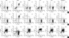
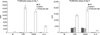
Whole B cells have been classified as B-1 and B-2 cells. Flow cytometry showed that almost all lymph node B cells had the B-2 phenotype, which included CD5low, CD23high, CD43low, CD80 (B7.1)low, CD86 (B7.2)low, and Syndecan-1low, similar to the phenotype in normal spleen (Fig. 1).
Lymph node B cells do not live longer
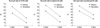
Upon stimulation, lymph node B cells showed 60±5.1% survival on day 2 and 25±3.2% survival on day 5, whereas unstimulated lymph node B cells showed 39±4.1% and 10±2.3% survival on days 2 and 5, respectively. These survival rates were no higher than those of splenic and peritoneal B cells (Fig. 2).
B cells of cervical lymph nodes and spleen secrete immunoglobulin in response to stimulation
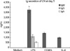
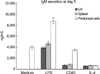
We found that unstimulated B cells from cervical lymph nodes and spleen did not spontaneously secrete immunoglobulin. These cells secreted IgM upon stimulation with LPS (3,300 ng/mL on day 3 and 4,286 ng/mL on day 5), CD40L plus anti-CD8 antibody (1,147 ng/mL on day 3 and 1,392 ng/mL on day 5) or IL-4 (1,017 ng/mL on day 5) (Fig. 3). In contrast, unstimulated peritoneal B cells secreted IgM, and secreted higher concentrations of IgM than either splenic or lymph node B cells (Fig. 4).
Stimulated cervical lymph node and splenic B cells enter S phase on day 2
Unstimulated B cells of cervical lymph nodes and spleen did not enter S-phase, as shown by incorporation of [3H] thymidine, after 24 hr in culture, but actively entered S phase after 48 hr of stimulation. These cells incorporated [3H] thymidine upon stimulation with soluble CD40L/CD8α (229×103±23×103 cpm at 24 hr and 3,526×103±307×103 cpm at 48 hr), LPS (256×103±25×103 cpm at 24 hr and 2,790×103±254×103 cpm at 48 hr), or IL-4 (171×103 ±18×103 cpm at 24 hr and 256×103±28×103 cpm at 48 hr) (Fig. 5). Although recombinant IL-4 was originally identified as a growth and differentiation factor for conventional B cells, it did not stimulate cervical lymph node and splenic B cells to enter S phase. Thus, it seems unlikely that cervical lymph node B cells are especially primed by antigenic stimulation to enter the cell cycle.
DISCUSSION
B cells are responsible for antibody-mediated immunity, which is also known as humoral immunity. These cells are not evenly distributed in the blood, bone marrow, spleen, thymus, and peripheral lymphoid tissues. Rather, the ratio of B cells to T cells varies by the tissue or organ. For example, human B cells are seldom found in the thymus and, in the blood, T cells are outnumbered by B cells by a ratio of 8:1. This ratio changes to 1:1 in the spleen, whereas B cells outnumber T cells by a ratio of 1:3 in bone marrow. We found that the ratio of B cells to T cells in murine cervical lymph nodes was 1:3.1 by negative selection methods and 1:2.5 by flow cytometry (1).
B cells have been classified as B-1 and B-2 cells. B-1 cells are distinguished from the more abundant conventional B (B-2) cells by expression of the pan-T cell surface glycoprotein, CD5. Additional identifying phenotypic characteristics of B-1 cells include surface Ig (sIg) Mhigh, sIgDlow, B220low, CD23low, and CD43high. In contrast, B-2 cells express Ig (sIg) Mlow, sIgDhigh, B220high, CD23high, and CD43low (3,9). We found that cervical lymph node B cells expressed CD5low, CD23high, CD 43low, CD80 (B7.1)low, CD86 (B7.2)low and Syndecan-1low, suggesting that these cells are comparable to B-2 cells.
We previously reported that splenic B cells are exclusively B-2 cells and peritoneal B cells are mainly B-1 cells (7). B-1 cells represent 10-25% of the B cells found in adult human peripheral blood and lymphoid organs. They are the principal lymphocyte population in the peritoneal cavity and represent a small proportion of splenic B cells, but they are absent from the peripheral blood in adult mice (2). Although B-1 (B220+ CD5+) cells have been reported to be absent from murine lymph nodes (10), we found that 3.5% of the B cells in murine cervical lymph nodes were B220+ CD5+ cells (1). Our phenotypic data, however, represent only a single point in time, and we cannot rule out the possibility that B-1 cells are generated in murine cervical lymph nodes and then depart.
When B cells are activated by T-dependent, T cell producing cytokines or T-independent mitogens, they proliferate or differentiate into antibody producing plasma cells or memory cells (11-13). After stimulation with antigen, B cells first produce IgM and IgD and thereafter switch to secrete IgG, IgA or IgE, a switching that follows a DNA recombination event (14). The class of Ig produced by B cells differs by organ sites, stimuli and/or pathophysiologic conditions. Thus, most of human B cells in the middle ear and nasal mucosa primarily secrete IgA, both under normal and pathologic conditions, whereas other B cells in these organs secrete IgG rather than IgM. In contrast, nasal polyps contain B cells capable of spontaneous, high-rate IgA secretion, followed by secretion of IgG and IgM (15,16). These findings suggest that the middle ear mucosa and nasal mucosa contain distinct immune systems, which exhibit features similar to those at other mucosal sites. For example, IgG producing adenoid CD5+ and CD5- B cells were most abundant in adenoids, followed by IgA-producing and IgM-producing cells (17).
We found that B cells in murine cervical lymph nodes secreted mainly IgM, followed by IgG and IgA, when stimulated with LPS, a T-independent mitogen; soluble CD40L/CD8α, a stimulus provided by T cells; or IL-4, a cytokine produced by T cells. These finding suggests that B cells in cervical lymph nodes are stimulated or respond differently than B cells at the nasal and middle ear mucosal surfaces. In contrast, our finding, that murine cervical lymph node B cells do not secrete any Ig spontaneously, was unexpected. This result suggests that cervical lymph node B cells may not be subjected to continual antigenic stimulation from the naso- or oro-pharynx. Rather, it is likely that these cells have the same characteristic features as splenic B cells.
There are several differences between B-1 and B-2 cells concerned with Ig secretion (2,7). In normal individuals B-1 cells are responsible for the production of most nonimmune serum IgM, as well as producing substantial amounts of resting IgA. B-1 cell-derived Ig often recognizes discrete microbial cell wall determinants, suggesting that these cells produce natural antibodies, representing a set of specificities encoded in the germline and evolutionarily retained. These natural antibodies provide, at low affinity, a degree of serological protection against a range of microorganisms prior to the immunization that accompanies microbial pathogenesis (18). Primary B cells are normally arrested in the G0 phase of the cell cycle (6,7,19,20). Following the crosslinking of antigen specific surface Ig receptors on B cells, a number of early metabolic changes occur, which eventuate in cell cycle progression and entry into S phase.
B-1 and B-2 cells also show different proliferation responses. We found that murine cervical lymph node B cells responded in vitro to LPS or CD40L/CD8α in terms of cell cycle entry. While they did not proliferate actively after 1 day, they proliferated at 2 days. We had conjectured that lymph node B cell would be longer lived than splenic B cells, due to continuous stimulation from the naso- or oropharynx; however, their survival rates, with or without stimulation, were not high, suggesting that murine cervical lymph node B cells have the characteristics of B-2 cells.
The phenotypic characteristics of nodal B cells include CD 5low, CD23high, CD43low, B220high, sIg Mlow, sIgDhigh, Mac-1low, CD80 (B7.1)low, CD86 (B7.2)low, and CD138 (Syndecan-1)low. Our finding, that 3.5% of B cells were B220+ CD5+ suggests that a small population of B-1 cells is present in cervical lymph nodes. Unlike our findings with peritoneal B cells, which spontaneously produce Ig and enter S phase after 1 day of stimulation, we did not observe these characteristics in cells from murine cervical lymph nodes. Despite our expectation that these lymph node B cells would be continually stimulated by antigens from the naso- or oro-pharyngeal mucosa, we found no evidence for unusual surface phenotype, differentiation, survival rate or proliferation. These results indicate that cervical lymph node B cells correspond to conventional B-2 cells and that they have similar characteristics to splenic B cells (B-2), but not to peritoneal B cells (B-1).
 XML Download