

 PDF
PDF ePub
ePub Citation
 Print
Print


INTRODUCTION
A santorinicele is a focal cystic dilatation of the terminal portion of the dorsal pancreatic duct at the minor papilla, and has been reported in patients with pancreas divisum (1). Clinically this rare anomaly has been suggested to be a possible cause of a relative stenosis of the minor papilla, which in association with pancreas divisum, results in a high intraductal pressure and resultant pancreatitis (1-7). We recently encountered a patient with a santorinicele without pancreas divisum. Although there is only one report of a santorinicele without pancreas divisum, it was combined with bifid pancreas, which is a congenital anomaly where the primitive pancreatic channels fail to fuse into a single main duct during the development of the pancreas (8). We describe a typical case of a santorinicele without pancreas divisum or other congenital pancreatic anomalies that was identified incidentally by multidetector row computed tomography (MDCT).
CASE REPORT
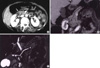
A 67-yr-old woman was admitted complaining of paraumbilical and lower left abdominal pain with intermittent hematochezia. She had been previously treated for diabetes mellitus and hypertension with an oral hypoglycemic agent and a calcium channel blocker for the previous 10 yr. She was a non-smoker, and non-drinker with no family history of gastrointestinal disease. The physical examination was negative except for the paraumbilical and left lower abdominal tenderness. The laboratory studies, including a complete blood count, liver function tests, serum amylase level, were within the normal range. The colonoscopic findings were diffuse erythema, edema, ulcer, and friability of the mucosa through the proximal descending and distal transverse colon, which was consistent with ischemic colitis. MDCT (LightSpeed Pro; GE Medical Systems, Milwaukee, WI, U.S.A.) was performed after the colonoscopy. The CT scan protocol was as follows: 150 mL of iopromide (Ultravist 370; Schering, Berlin, Germany) was injected through an 18-gauge angiographic catheter that was inserted into a forearm vein at a flow rate of 3 mL/sec by using a mechanical injector. Portal venous phase images were acquired following a scanning delay of 70 sec from the time of initiation of contrast material injection, and the delayed phase images followed after a scanning delay of 3 min. The CT parameters included a detector configuration of 1.25 mm×16, a gantry rotation speed of 0.5 sec, and a table feed of 20 mm per gantry rotation. CT showed mild and diffuse wall thickening from the distal transverse colon to the descending colon. It also showed a small round low-density lesion at the distal dorsal pancreatic duct, which was adjacent to the duodenal wall (Fig. 1A). The 1.25-mm transverse CT scans were reconstructed at 0.625-mm intervals on a dedicated workstation (Advantage Window 4.2; GE Medical Systems). Reformation was performed using a minimum intensity projection technique. The coronal reformatted image using a minimum intensity projection technique clearly showed a cystic dilatation of the distal dorsal pancreatic duct and a communication between the ventral and dorsal pancreatic ducts (Fig. 1B). These findings were compatible with a santorinicele without pancreas divisum. The dorsal pancreatic duct was equal to or slightly more prominent than the ventral pancreatic duct. The subsequent magnetic resonance cholangiopancreatography (MRCP) confirmed the cystic dilatation of the distal dorsal pancreatic duct. There was no evidence of pancreas divisum or any other congenital anomaly of pancreas (Fig. 1C). Based on the findings of imaging studies, the diagnosis of a santorinicele without pancreatic divisum was made.
The patient did well after supportive cares including intravenous fluids and antibiotics for ischemic colitis, and then was discharged on the 9th day of admission without any problems.
DISCUSSION
A santorinicele is believed to be analogous to a dilatation of the most distal common bile duct, which is commonly known as a choledochocele. It was first described in 1994 by Eisen et al. (1), who reported 4 patients with pancreatitis and pancreas divisum accompanied by a focal cystic dilatation of the terminal portion of the dorsal pancreatic duct by endoscopic retrograde cholangiopancreatography (ERCP). Since the first description, santoriniceles have been reported in patients with pancreas divisum, either complete or incomplete (2-7). Most reports of santoriniceles have common features such as pancreas divisum and pancreatitis. The santorinicele has been suggested to be a possible cause of the relative stenosis of the accessory papilla, which in association with unfused dorsal and ventral ducts results in the high intraductal pressure responsible for the recurrent episodes of acute pancreatitis. Therefore, a sphincterotomy or balloon dilatation is believed to be effective in patients with a santorinicele and pancreatitis (3-5, 7).
The prevalence of this rare anomaly is unknown, and it is unclear if it is congenital in origin or is an acquired lesion secondary to a stenosis of the dorsal duct orifice. Because most santoriniceles have been reported in elderly patients, it has been assumed that a santorinicele is most probably an acquired rather than a congenital condition. Some santoriniceles are associated with an adjacent duodenal diverticulum. Structural changes might contribute to the acquired mucosal weakness, thereby facilitating the formation of a santorinicele. The association with a duodenal diverticulum may further support the acquired etiology. However, a report of a santorinicele in a pediatric patient without a duodenal diverticulum suggests that the pathogenesis is congenital in some cases (4).
Since all santoriniceles reported were detected in patients with pancreatic divisum, it was unclear if a santorinicele might be present in patients without this anomaly. In 2003, a santorincele without pancreatic divisum was reported by Byeon et al. (8). This is the only report of a santorinicele not associated with pancreas divisum. However, instead of pancreas divisum, the patient had another congenital anomaly, bifid pancreas, which is an anatomical variant where the main pancreatic duct is bifurcated along its length. On the other hand, our case was characteristic in two ways. First, the patient did not have pancreas divisum and other congenital pancreatic anomalies. Second, CT detected the santorinicele incidentally. Since all santorinicele reported were diagnosed by MRCP or ERCP, this is the first case reported of a santorinicele without pancreas divisum detected by CT.
In conclusion, this case demonstrates that a santorinicele can exist without pancreas divisum. Furthermore, MDCT is believed to be a very useful diagnostic technique for delicate lesions like as the present case.
 XML Download