

 PDF
PDF ePub
ePub Citation
 Print
Print


INTRODUCTION
It is well known that the success rate for traditional trabeculectomy is much lower in patients with intractable glaucoma resulting from iris neovascularization or uveitis, or in patients with profuse subconjunctival fibrosis due to any previous ocular procedure including filtering surgery (1). In these patients, aqueous drainage devices have come into use to preserve the visual function and to relieve pain caused by high intraocular pressure (2). Among several devices in use, Ahmed glaucoma valve implant has become one of the most popular devices because it lowers intraocular pressure satisfactorily with low incidence of postoperative hypotony. But the exposure of the some parts of the valve is noted occasionally and bullous changes of corneal epithelium can occur infrequently due to the dysfunction of endothelial cells with the most troublesome symptoms being decreased vision and severe ocular pain.
In an effort to decrease incidence of implant exposure and to alleviate ocular pain, we utilized amniotic membrane over the tubal portion of the valve during Ahmed valve implantation and placed an amniotic membrane graft over the cornea.
The purpose of our study is to investigate if these new applications of amniotic membrane used in conjunction with Ahmed valve implantation can be useful in stabilizing ocular surface and in lowering postoperative complications, thus relieve severe ocular pain in the long run.
MATERIALS AND METHODS
We retrospectively reviewed the medical records of sixteen patients with sixteen eyes who were followed in our clinic from February 2002 to March 2004. Of sixteen patients, ten patients were male and six were female. The mean age was 66.1±4.3 yr, two patients had the history of detachment of retina, two had experienced trauma, five were pseudophakic, two were aphakic, and one had received both penetrating keratoplasty and intraocular lens insertion. The mean of intraocular pressure checked before the operation was 43.9±9.0 mmHg. They had undergone Ahmed valve insertion with folded amniotic patch over the implant, and debridement of corneal epithelium combined with amniotic membrane graft over the exposed stroma for the control of the intractable glaucoma with bullous keratopathy. With informed consent and approval by the Institutional Review Board, human placenta was obtained shortly after Cesarean delivery. The amniotic membrane was harvested and preserved using the method described by Kim and Tseng (3).
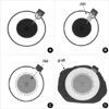
The drainage device used in this study was Ahmed type. This pear shaped polypropylene device was implanted into eyes under retrobulbar anesthesia. All procedures were performed by one surgeon. First, a fornix based conjunctival flap was created in superior-temporal quadrant. Tenon's capsule was incised and the tissue was dissected off the sclera to create a pocket for the plate of the valve. Care was taken to avoid injury to any adjacent extraocular muscles. The tube was irrigated with balanced salt solution to ascertain that the valve functions normally. The body plate of the implant was inserted into the pocket. It was placed 8-9 mm posterior to the corneal limbus and secured to the sclera with 10-0 nylon. The anterior chamber was entered parallel to the iris plane just posterior to the limbus using 23G needle. The tube was trimmed so that the cut bevel faced the corneal endothelial surface and was inserted into the anterior chamber along the previously created needle track, making sure the cut edge does not extend over 2 mm from the entry site. The remaining portion of tube was overlaid with amniotic membrane patch graft. The amniotic membrane was cut into 6×18 mm rectangular piece and folded in half so that stromal surfaces face each other, and then the membrane was secured to the sclera using 10-0 nylon interrupted suture. The conjunctiva and Tenon's flaps were sutured back to the limbus. The bullous corneal epithelium was scraped off mechanically using microsurgical sponge, fine forceps, and Beaver blade. Peripheral corneal epithelium within 1 mm of limbus was not removed. Then an amniotic membrane was cut into a disk shape and was placed on the cornea with the stromal surface down. The membrane was secured to the eye with 10-0 nylon in a continuous manner. Finally, we covered the whole surgical field using a large amniotic membrane with amnion side down as a temporal patch graft and secured it sclera with 10-0 nylon interruptedly (Fig. 1). All the eyes received topical steroids and antibiotics four times a day. At each follow up visit, visual acuity, intraocular pressure, and postoperative conditions including persistence of pain or occurrence of complications were checked carefully.
RESULTS
The mean follow up period was 8.4±3.2 months. The mean intraocular pressure (IOP) was significantly dropped from 43.9±9.0 mmHg to 11.3±3.4 mmHg on the first postoperative day and 16.1±1.8 mmHg at the last follow up (p<0.01 by paired t-test). It was relatively well controlled throughout the follow-up periods. Neither hypotony nor transient hypertensive period was noted. Vision was not improved much except in one patient due to the severity of preexisting pathologies. The corneal epithelium recovered well without any bullae in all patients, and the averaged time elapsed for the full reepithelialization of the cornea was 10.1±1.3 days. We could not find any evidences of tubal exposure or erosion of conjunctiva. The most notable finding was that the pain vanished in all patients as the corneal surface became stabilized rapidly with the use of amniotic membrane even in presence of late partial dissolution of amniotic membrane and recurrent bullae in some cases (Table 1). None of the patients experienced severe ocular pain after surgery, and the IOP was significantly lowered by the last follow up (p<0.01 by paired t-test).
The following three cases demonstrate well that our new application of amniotic membrane during Ahmed valve implantation reduced preoperative IOP and relieved pain significantly without any serious complications.
Case 1
A 63-yr old man visited our clinic complaining of left ocular pain that had started 7 days before. He had lost his left vision due to optic neuropathy after a traffic accident. He had no light perception in that eye, and his IOP was measured as high as 35 mmHg. On the slit lamp examination some corneal opacities associated with epithelial bullae were noted, along with intrastromal edema and neovascularization on corneal surface. Surgery was performed as aforementioned method.
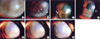
The IOP measured after surgery was 12 mmHg. Full reepithelialization of cornea was achieved by the fifth postoperative day. At his last visit IOP was well controlled at 18 mm Hg, and no complications associated with the valve have occurred. Cornea reepithelialized, and no bullae were found. He has never experienced any ocular pain subsequently even though focal melting of amniotic membrane developed (Fig. 2A, B).
Case 2
A 61-yr old man who had received cataract extraction surgery 7 yr before, visited our clinic due to recurrent ocular pain which has become increasingly more severe with time. He could not perceive light and his IOP was 42 mmHg. The slit lamp examination revealed a large bulla with opacity and stromal edema in the inferior portion of cornea. He received the same operation sequentially. The IOP after surgery was 15 mmHg. The full reepithelialization of cornea was achieved by the sixth postoperative day. IOP was well controlled and remained under 20 mmHg through his last visit and no complications associated with the tube were noted. Cornea epithelium looked stabilized. He did not complain of ocular pain thereafter (Fig. 2 C, D).
Case 3
A 64 yr old woman who had received vitrectomy with silicon oil replacement due to retinal hemorrhage and detachment 7 yr before, visited our clinic due to the severe right ocular pain, which had developed 1 week ago. The slit lamp examination revealed a number of bullae with almost total corneal opacification and stromal edema. She could not perceive light and her measured IOP was 35 mmHg. The scheduled operation was done as described before. The IOP measured after the surgery was 11 mmHg and was well controlled measuring as low as 18 mmHg on her last visit after 10 months postoperatively, and no complications associated with the tube were found. The cornea epithelium was well established and no bullae were found. The ocular pain disappeared after that (Fig. 2E-G).
DISCUSSION
Since the drainage device was first used in 1912, various kinds of devices have been developed (4-7). Ahmed valve implant has many advantages over other devices owing to its large drainage surface and less incidence of hypotony. But the most common complication still not solved is the erosion of conjunctiva that covers the implant and as a consequence, the exposure of some parts of the implant itself (7, 8). Moreover the exposure of implant tends to recur and can lead to secondary bacterial infection, which can spread into intraocular structure and can result in devastating endophthalmitis. In such cases the implant has to be removed eventually. For this reason, it is essential to carefully cover the tube and plate with a graft during surgery. The graft used for this purpose should be stable enough not to be absorbed easily and should not impose any pressure on adjacent structures. Most commonly, a human donor scleral patch graft has been used to cover the tubal portion of implant. However, even with the best efforts, the exposure of the implant continues to occur, and many other materials have been tried as a substitute for human donor scleral patch (9-12). Even though, some of these materials are difficult to obtain, and there has been a concern that human dura mata may transmit prion diseases such as Creutzfeldt-Jakob. Therefore, there have been continued efforts to find more suitable graft materials.
Bullous keratopathy are caused by the loss of endothelial function and this in turn increases stromal hydration and brings about loss of keratocytes, further hydration into intraepithelial space (13, 14). The patients may suffer from severe ocular pain and decreased vision regardless what had initiated the insult to the cornea. Many modalities of the treatment have been considered. In patients with reasonable vision potential, the treatment of choice is corneal transplantation. But in patients with limited vision potential, the bandage contact lens, anterior stromal puncture, annular keratotomy, therapeutic photorefractive keratectomy, or conjunctival flap can be considered as a surgical option to relieve pain and to support corneal surface (15-18). Among these options, conjunctival flap has a long history of wide use and is a well proven and time-honored method for restoring ocular surface integrity in diseased cornea. However poor cosmetic results and difficulty in examining ocular pathology post-operatively have been the major disadvantages of conjunctival flap. Recently amniotic membrane was used to treat symptomatic bullous keratopathy with excellent results (19).
Amniotic membrane is the innermost part of the placenta. Since the study by Kim and Tseng on application of amniotic membrane to rehabilitate severely damaged cornea in 1995, its indications for use have been expanded rapidly (20-24). Todays, the use of amniotic membrane as a carrier for epithelial cells expansion is under investigation. The exact mechanisms for how amniotic membrane induces favorable responses in many diverse conditions are not fully understood, nor are the membrane's compositions. What is known about amniotic membrane so far is that it contains abundant extracellular matrix materials such as fibronectin, laminin, type IV collagen and integrin, as well as various protease and growth factors. So amniotic membrane acts as a mechanical barrier in many conditions in which ocular structural reinforcement is needed, and thereby, it lessens ocular pain in exposed cornea that has ocular surface pathology and offers substrate for epithelial migration. By affecting down regulation of TGF-β system, amniotic membrane can also help corneal cells to differentiate normally and retain their cellular characteristics and thus prevent scar formation and maintains corneal clarity (25). The protease inhibitors may play some roles in restoring normal ocular architecture (26).
The advantages of amniotic membrane over other materials are that it is easy to obtain, is relatively cheap, and is easy to manipulate. Also it does not induce rejection. When needed, the thickness of graft can be increased substantially by folding the membrane. Moreover, since it is semi-translucent, it renders cosmetically superb result and readily allows examination of the underlying structure, thus enabling early detection of any complications.
With these backgrounds, we performed Ahmed valve insertion with amniotic membrane patch graft over the implant itself, and debridement of corneal epithelium with amniotic membrane graft over the exposed stroma as a single operation in patients with painful bullous keratopathy secondary to the intractable glaucoma. The amniotic tissue transplanted over the tube and plate portion of the implant remained intact until the last follow-up and no complications associated with the exposure of the implant were noted. After the implantation of drainage device, a transient rise of IOP is often seen during the first 3 months postoperative period. This period of transient rise in IOP is called hypertensive period (8). In our study, however, we could not detect hypertensive phase in any of the patients, and the IOP was well controlled throughout the study. As the hypertensive period is thought to be caused by the formation of fibrotic bleb around the implant, the role of amniotic membrane in reducing fibrosis may play some roles in preventing IOP rise. Our study showed excellent results in the control of intraocular pressure: the mean preoperative IOP was 43.9±9.0 mmHg and it dropped significantly to 16.1±1.8 mmHg at the last visit. The IOP of all patients were controlled within the desirable range until the end of this study independent of the number of antiglaucoma medication in use. These results were better in comparison with those reported in the earlier studies (8). And moreover, none of the patients required secondary procedures such as needling of bleb and injection of 5-fluorouracil to control the IOP.
In the management of bullous keratopathy, amniotic membrane is easy to manipulate during surgery does not impair limbal cell function. Intolerable pain disappeared in all patients immediately after the procedure. And we experienced prominent recovery of vision in one pseudophakic patient. Her vision improved from light perception preoperatively to 0.4 at the last follow up. The translucency of the amniotic membrane may play some roles in this improvement, thus lessen the need of penetrating keratoplasty in the future.
We noted partial dissolution of the amniotic membrane and recurrence of small bullae in some patients, but none of these patients reported any discomfort during the follow-up periods. It is not easily explained how the relief of ocular pain was maintained in such cases. As the mean follow up period was relatively short, we have to investigate further in what ways amniotic membrane have contributed in this regards.
In summary, amniotic membrane transplantation is effective way to provide support for Ahmed valve and is an efficacious treatment for bullous keratopathy as it stabilizes ocular surface rapidly.

 XML Download