

 PDF
PDF ePub
ePub Citation
 Print
Print


INTRODUCTION
Heat shock proteins (HSP) are highly conserved cellular stress proteins present in every organism from bacteria to man. It was first observed in 1962 that the salivary gland chromosomes of the fruit fly, Drosophila melanogaster, exhibited a characteristic puffing pattern after exposure to heat (1).
HSP act under physiological condition as molecular chaperones which are involved in mediating the folding and transport of other intracellular proteins and in some cases their assembly into oligomeric structures. HSP act as chaperones by participating in assembly of proteins without being part of the final protein structure (2). They are induced in response to cellular stress which includes changes in temperature, the presence of free oxygen radicals, viral and bacterial infections, heavy metals, ethanol and ischemia (3).
HSP production is enhanced during in vitro embryo culture and they are among the first proteins produced during mammalian embryo growth and presence or absence of HSP influences various aspects of reproduction in many species.
HSP are classified into different families according to their molecular weight, the 27, 60, 70 and 90 kDa HSP. HSP 60 and HSP 70 are most important in the field of reproduction. HSP 60 is located in the cell surface (4), mainly in the mitochondria (5, 6). HSP are immunodominant antigens of microbes such as Chlamydia trachomatis, which have been recognized as the main cause of tubal infertility. Many couples with fertility problems have had a previous genital tract infection, have become sensitized to microbial HSP and a prolonged and asymptomatic infection may trigger immunity to microbial HSP epitopes that are also expressed in man (7). Antibodies to both chlamydial and human HSP are found at high titre in sera and hydrosalpinx fluid and in a mouse embryo culture model (8), these antibodies impaired the mouse embryo development at unique developmental stage.
Numerous studies have demonstrated the beneficial effects of cellular monolayer of various somatic cell types on mammalian embryonic development (9-11). Many different somatic cell types have been used for coculture, including human oviductal epithelial cells (9), human endometrial fibroblasts (12), bovine oviductal epithelial cells (9). However, the use of human or animal cell lines for the coculture of embryos pose many problems such as viral infections. Vero cells, derived from African green monkey kidney, share a common embryonic origin with cells from genital tracts. However in addition, they are potentially safe to use since they are highly controlled for viruses and other contaminants. Therefore coculture using Vero cell has been widely utilized to enhance embryo viability and development (13).
The aim of this study was to investigate the detrimental effects of HSP 60 on the development of mouse embryos in vitro and to demonstrate whether Vero cells overcome these adverse effects.
MATERIALS AND METHODS
Superovulation and collection of embryos
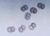
5-6 week female mice were superovulated with injection of 7.5 I.U. pregnant mare's serum gonadotropin (PMSG; Sigma Chemical Co., St. Louis, MO, U.S.A.), followed 48 hr later by 7.5 I.U. human chorionic gonadotropin (PMSG; Sigma). After mating overnight, females exhibiting vaginal plug were killed 48 hr post hCG administration and 2-cell embryos were flushed from the oviducts with Dulbecco's phosphate buffered saline (D-PBS, Gibco BRL, N.Y., U.S.A.) supplemented with 0.4% bovine serum albumin (BSA, Sigma) (Fig. 1).
Preparation of Vero cell monolayer
From the frozen cells, flasks were washed with Ham's F-10 (Gibco BRL)+10% FBS and Vero cells, 2-3×10⁶ per well were cultured in tissue culture flask (75 cm2, Falcon) in a 5% CO2 humidified air incubator. After trypsinization, the cell suspension was divided into three parts. One third was frozen, another one third was used to seed again in a new flask, and the remaining portion was used to seed wells at a concentration of 105 cells per well reaching confluence within 3 days. The cells must not be passaged repeatedly because the growth is significantly reduced after four subpassages.
Embryo culture
Ham's F-10 supplemented with 10% FBS was used as culture medium in the control group. The 2-cell embryos in culture medium supplemented with 100 µg/mL of monoclonal antibody specific for mammalian HSP 60 (StressGen, Canada) with or without Vero cells were incubated at 37℃ in a 5% CO2 humidified air chamber respectively and we observed mouse embryo development every 24 hr for 3 days.
RESULTS
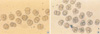
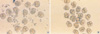
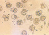
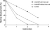
At day 1, 45% of the embryos cultured in Ham's F-10 plus anti-HSP 60 with Vero cells developed to 4- to 8-cell stage as compared with 29.2% of those without Vero cells (Table 1) (Fig. 2). Similarly at day 2, morulae were well observed in 22.1% of cultures with Vero cells as opposed to 2.7% of those without Vero cells (Table 1) (Fig. 3). In blastocystic process, however, the beneficial effects of Vero cells were not noted (Table 1) (Fig. 4).
DISCUSSION
The spontaneous expression of HSP as an essential part of embryo development is well documented and the presence or absence of HSP influences various aspects of reproduction in many species. It was reported that 2-cell mouse embryos were cultured in the presence or absence of monoclonal antibodies specific for mammalian HSP 60, HSP 70 and HSP 90 and these antibodies impaired the mouse embryo development at unique developmental stages (14). Our data in the previous study showed that anti-HSP 60 elicited a strong inhibitory effect on mouse embryo growth (15).
The detrimental effects of HSP on reproduction are based on several mechanisms. Firstly, HSP can induce a persistent inflammatory response (16). Secondly, HSP molecules serve as antigenic targets for the immune system (7). Finally, the extensive amino acid sequence homology between human and microbial HSP could result in autoimmune mediated reproductive failure (17).
The Chlamydia trachomatis infection is common and the major cause of infertility due to occluded Fallopian tubes. During the course of an acute Chlamydia trachomatis infection, immunity is restricted to HSP 60 epitopes that are specific to the invading micro-organism. Most patients with infertility problems due to tubal occlusion have experienced a chronic persistent chlamydial infection (7). The cells chronically infected with C. trachomatis synthesize only low levels of structural components but continue to produce chlamydial HSP 60 at high levels (18). Thus, some women with asymptomatic and untreated C. trachomatis infections and tubal infertility have experienced a long-term exposure to chlamydial HSP 60. Since chlamydial and human HSP share extensive amino acid sequence homology (17), it has been proposed that a prolonged exposure of the immune system to chlamydial HSP 60 and a concomitant exposure to both the chlamydial and human HSP 60 may lead to autoantibody formation (7).
Human (host) HSP 60 expression is physiologically expressed during pre- and peri-implantation stages by the embryo and the maternal deciduas. Host HSP 60 expression in early pregnancy reactivates lymphocytes previously sensitized to chlamydial HSP 60. The activated lymphocytes release proinflammatory cytokines, which induce also other lymphoid cells to release inflammatory and cytotoxic mediators. Cellular and humoral immune system activation disturbs immune regulatory mechanisms necessary to implantation and maintenance of embryos. Antibodies to HSP impair embryo development as our previous study was shown (15). Embryos are less protected from adverse environmental conditions and are more likely to degenerate or undergo apoptosis.
The mechanism of anti-HSP60 related inhibition of mouse embryo development in vitro is completely unknown. The zona pellucida of mouse oocytes and zygotes is permeable to macromolecules. Molecules up to 170 kDa have been shown to penetrate through the zona pellucida of post-ovulated mouse oocytes (19). In addition the permeability of mouse zona pellucida to IgG with induction of embryo damage has been demonstrated (20).
It is now generally accepted that Vero cells provide beneficial effects on improving embryo quality in many animal species. Cocultured embryos on the Vero cell monolayer have higher cell numbers and less fragmentation during cleavage than conventionally cultured ones (21). In addition, Vero cells have been shown to provide significant improvements in morphology and cleavage embryos (22). The resulting embryos, therefore, are more likely to implant and develop progressively.
Somatic cells such as Vero cells may provide beneficial effects on embryo development by manufacturing an in vitro environment that is compatible with normal embryo development (23). However, it is unclear why the coculture with Vero cells improves embryo development. Some claim that they remove toxic compounds from the culture medium, such as heavy metal divalent cations and metabolic inhibitors (11, 24), but others support the hypothesis that various soluble factors, including mitogenic factors secreted by Vero cells, are more important for better embryos development, although their physiologic roles are yet to be determined (10, 23).
Serum proteins decrease embryotoxicity in the conventional embryo culture system by chelating toxic components in the media (25). The fact that coculture in a simple serumfree medium exerted beneficial effects on embryos suggests that toxic substances were removed by the cells in culture (26). Hypoxanthine, which is commonly found in most good culture medium formulations, has been shown to induce the 2-cell block in mouse embryos and this agent can be removed by the somatic cells to overcome the block (27). All these observations suggest that Vero cells seem to function by removing the inhibitory substances in media rather than by producing specific factors that enhance embryonic development. We found that Vero cells are likely to overcome the detrimental effect of anti-HSP 60 to some degree (Fig. 5).
We suggest that Vero cell coculture will promote reproductive outcome in patient previously sensitized to microbial (e.g. Chalmydia trachomatis) HSP 60 which is common cause of female infertility.
 XML Download