

 PDF
PDF ePub
ePub Citation
 Print
Print


INTRODUCTION
Posttransplant lymphoproliferative disorder (PTLD) is a well-recognized complication of solid organ and hematopoietic stem transplantation (HSCT), which is usually associated with Epstein-Barr virus (EBV). PTLD represents a heterogeneous group of abnormal lymphoid proliferation, generally B cells, which occurs in the clinical setting of T-cell dysfunction (1-3). The incidence of PTLD after solid organ transplantation (SOT) ranges from 2% in renal transplantation to 30% in intestinal and multiorgan transplantation. PTLD has been rare following allogeneic HSCT, but intensive immunosuppression, such as T-cell depletion of grafts, or antithymocyte globulin (ATG), to prevent and treat graft-versus-host disease (GVHD), can markedly increase the risk (1, 3-5). An underlying immunodeficiency and the use of alternative donors, such as matched unrelated donors or HLA-mismatched family members, are additional risk factors for the development of PTLD (1, 3, 4). During the past decade, the use of alternative donors has increased and this has placed recipients at a high risk for GVHD and consequent intensive immunosuppression.
As a result of these changes, PTLD has been encountered more frequently than in the past. We observed the incidence, as well as the clinical and pathologic features of PTLD among 1,116 patients who underwent allogeneic HSCT at Catholic HSCT Center in Korea.
MATERIALS AND METHODS
Study population
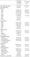
From January 1995 to December 2003, a total of 1,116 adult patients received allogeneic HSCTs at the Catholic HSCT Center. Acute GVHD was assessed and graded according to previously published data (6). In most cases, acute GVHD was treated with methylprednisolone pulse therapy alone. In the corticosteroid-resistant cases, ATG was used as the second-line treatment, or another immunosuppressant, such as mycophenolate mofetil or tacrolimus, was added. Acyclovir (800 mg/day) was given orally from day 7 to engraftment and was followed by long-term low dose (200 mg/day) oral administration for 6 months posttransplant. For cytomegalovirus (CMV) prophylaxis, high dose acyclovir (10 mg/kg three times daily) was given intravenously to patients with matched unrelated donor (MUD) HSCT, and ganciclovir (5 mg/kg twice daily) was given intravenously to patients with family mismatched (FMM) HSCT. Seven cases of PTLD were identified among 1,116 allogeneic HSCT recipients. We reviewed retrosepctively the medical and pathology records of these 7 patients. The details of the study population and cases of PTLD are summarized in Table 1.
Pathology
Biopsy specimens were obtained from lymph nodes in 4 cases, an oropharyngeal mass in 1 case, gastric tissue in 1 case, and colonic tissue in 1 case. Diagnoses were established through clinicopathologic correlation using routine histology, immunohistochemistry, and detection of EBV by in-situ hybridization for Epstein-Barr early RNA (EBER) by the method described previously in the literature (7).
RESULTS
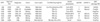
Seven patients developed PTLD following allogeneic HSCT: 4 MSD, 2 MUD, 1 full haplotype mismatched. The overall incidence of PTLD was 0.6% (7/1116). None was diagnosed postmortem. The median age was 37 yr (range, 17-45 yr) and the male to female ratio was 6:1, which demonstrated a predilection for males. All the patients, except one who underwent a full haplotype mismatched HSCT, were matched at the HLA A, B and DR loci. The underlying hematologic diseases were: chronic myeloid leukemia (CML) in three patients, acute myeloid leukemia (AML) in two, myelodysplastic syndrome (MDS) in one, and severe aplastic anemia (SAA) in one. For the 4 patients who underwent MSD HSCT, conditioning regimens were: total body irradiation (TBI) (1,200 cGy fractionated irradiation) plus cyclophosphamide (60 mg/kg/day for 2 days) in two patients; TBI (1,320 cGy fractionated irradiation) plus busulfan (3.2 mg/kg/day intravenously for 2 days) in one; and procarbazine (6.25 mg/kg for 6 days) plus cyclophosphamide (50 mg/kg for 4 days) plus ATG (Thymoglobuline, Sangstat, France, 1.25 mg/kg for 3 days) in one patient with SAA. Two patients who underwent MUD HSCT received a conditioning regimen of TBI (1,200 cGy) plus cyclophosphamide (60 mg/kg/day for 2 days). One patient who underwent haploidentical HSCT, received TBI (1,200 cGy), fludarabine (40 mg/BSA/day for 4 days), busulfan (3.2 mg/kg/day intravenously for 3 days) and ATG (Thymoglobuline, Sangstat, France, 1.25 mg/kg/day for 4 days), followed by donor-derived T-cell depleted peripheral stem cells by CliniMACS for 4 consecutive days. GVHD prophylaxis consisted of cyclosporin A with a short course of methotrexate in five patients, tacrolimus with a short course of methotrexate in one, and no GVHD prophylaxis was provided to one patient with haploidentical HSCT. Acute GVHD occurred in 5 patients: one patient with grade I, two with grade II, and two with grade III. All of them were treated with steroids and demonstrated good responses to steroid therapy alone. EBV serology prior to HSCT was available and was seropositive in all of the recipients. All recipients and donors were CMV seropositive. The characteristics and transplantation details of the 7 patients with PTLD are summarized in Table 2.
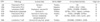
According to the WHO classification (8), 3 cases of PTLD were polymorphic, one was plasmacytic hyperplasia, and 3 were monomorphic, of which two were diffuse large B-cell lymphoma and one was large T-cell lymphoma. In situ hybridization for EBER was positive in 4 cases, negative in 2 cases and not performed in one case. The pathologic results are summarized in Table 3.
Symptoms and signs related to PTLD developed at a median time of 130 days (range, 83-244) after transplantation. The most frequently observed clinical finding was cervical lymphadenopathy (71.4%, 5/7), but clinical symptoms varied depending on the involved sites. Three patients had disseminated disease and four had localized disease. The first patient with disseminated disease (UPN1534) was admitted to the hospital with fever, chills, sore throat and cervical lymphadenopathy. He experienced acute grade III GVHD, which occurred on the 25th posttransplant day and responded to methylprednisolone pulse therapy. After that, there was no chronic GVHD and he was maintained on cyclosporin A (500 mg/day) and prednisolone (5 mg/day), but he was readmitted with high fever, chills, sore throat and cervical lymphadenopathy. A tonsillar biopsy confirmed polymorphic PTLD (lymphoma, polymorphic B-cell type) on the 91st posttransplant day. PTLD regressed partially after discontinuation of immunosuppressants and administration of high dose acyclovir. However, MDS relapsed and the patient died of a pulmonary hemorrhage on the 268th posttransplant day. The second patient with disseminated disease (UPN1907) presented with multiple and extensive lymphadenopathy. He had been treated with methylprednisolone (125 mg/day) due to acute grade II GVHD and demonstrated a good response. He had no chronic GVHD and was treated with cyclosporin A (300 mg/day) at the time of the diagnosis of PTLD. PTLD (diffuse large B-cell lymphoma) was confirmed by biopsy of cervical lymph nodes on the 130th posttransplant day. Treatment consisted of discontinuation of cyclosporin A, and initiation of high dose acyclovir and donor lymphocyte infusions (DLI, cell dose; 1×10⁶ CD3 cells/kg). PTLD did not regress but was stable after therapy. However, GVHD was aggravated after DLI and the patient died of pneumonia during the treatment of GVHD. The third patient (UPN 1985) underwent full haplotype mismatched HSCT with aggressive T-cell depletion and experienced CMV antigenemia just before the development of PTLD. He presented with generalized lymphadenopathy, diffuse lung infiltrates, and hepatosplenic involvement. Lymph node biopsy revealed diffuse large T-cell lymphoma with TCR-γδ positivity and positive in situ hybridization for EBER. Hepatic GVHD occurred concurrently with PTLD. He deteriorated so rapidly that there was no time for treatment with DLI and he died of progression of PTLD 8 days after the diagnosis. Four patients had localized disease. The involved sites were the stomach, colon, oropharynx with cervical lymph nodes, and cervical nodes alone. Three patients experienced acute GVHD (grade I in UPN426 and UPN1268, grade III in UPN1530) and responded well to steroid therapy, and only one of these patients (UPN1530) developed chronic GVHD (limited type) and was treated with tacrolimus (1 g/day) and prednisolone (20 mg/day). Two patients had polymorphic PTLD and one had plasmacytic hyperplasia, which was an early lesion with a good prognosis. One patient without acute GVHD (UPN1420) developed chronic GVHD concurrently with PTLD (diffuse large B-cell lymphoma). Treatment included discontinuation of immunosuppressants and DLI (cell dose; 5×10⁵ CD3 cells/kg in UPN426, 1×10⁶ CD3 cells/kg in UPN1268 and UPN1420) with or without high dose acyclovir in 3 patients; and one of the patients (UPN1530) with plasmacytic hyperplasia, had immunosuppressants discontinued and received high dose acyclovir alone. All four patients with localized disease demonstrated complete responses to therapy. Four patients are still alive and free of disease.
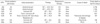
As described above, the response to treatment was relatively good (overall response rate; 71.4%, 5/7) and patients who had localized or polymorphic PTLD had better outcomes than those who did not. The median number of days of follow-up after diagnosis of PTLD was 831 (range, 8-2,750 days). Three patients died (overall mortality rate; 42.8%), but only one patient's death was directly attributable to PTLD (PTLD-related mortality rate; 14.2%). The clinical features and outcomes of the patients with PTLD are summarized in Table 4.
DISCUSSION
PTLD is a life threatening disease that encompasses a heterogeneous group of lymphoproliferative disorders ranging from reactive, polyclonal hyperplasia to aggressive non-Hodgkin's lymphoma (1, 3, 4). PTLD is a well-known complication following solid organ transplantations and its characteristics, as well as the strategy for management following the diagnosis, have been published extensively (3, 9-11). However, PTLD in the setting of HSCT, has not been reported in Korea. The purpose of this study was to examine the clinical and pathologic characteristics and outcomes of PTLD following allogeneic HSCT.
The incidence of PTLD after allogeneic HSCT has been reported to be approximately 1% (4, 5, 11). In this study, the overall incidence of PTLD of 0.6% (7/1116) was slightly lower than that of previous studies. However, a recent study reported that the incidence of PTLD following allogeneic HSCT was as high as 7.4%. In that study, all the patients with PTLD had acute GVHD and most of them were treated with ATG, which suggested that the occurrence of PTLD was strongly associated with the intensity of the immunosuppressive treatment (13). In our study, however, it appeared that PTLD was not as strongly associated with the grade of GVHD. Acute GVHD was not severe enough to require that patients undergo profound immunosuppression and patients responded well to steroid therapy alone. However, it should be taken into consideration that over 60% of the cases of PTLD were diagnosed postmortem in the previous studies in western countries; and by contrast, in this study there were no patients with confirmed cases of PTLD at autopsy, because the autopsy rate is very low in Korea. Therefore, it appeared that the incidence of PTLD and the role of immunosuppression in the development of PTLD were underestimated in this study compared to the previous reports (11-13). Furthermore, since transplants from alternative donors, which are complicated by the development of GVHD and a delay in immune reconstitution, have been increasing, the incidence and severity of PTLD is expected to rise in the future.
The majority of monomorphic PTLDs are of B-cell origin. In the SOT setting, approximately 13% of PTLDs are of T-cell origin and 20% to 38% of those are EBV positive (3, 14-17). However, in the HSCT setting, T-cell PTLD is very rare and only 5 cases have been reported; and only one case of PTLD following autolous HSCT proved to be associated with EBV (18-20). We observed one case of large T-cell lymphoma that proved to be associated with EBV by positive in situ hybridization for EBER. To our knowledge, this would be the first report of EBV-positive T-cell PTLD following allogeneic HSCT. Recently, Chuhjo et al. proposed the possibility that EBV can simultaneously infect B and T cells and can induce clonal proliferation of both lymphocytes in severely immunocompromised patients (21). Taken together, EBV may play a certain role in the development of T-cell PTLD after HSCT, although its pathogenesis has not been established. We think that aggressive immunosuppressive therapy in combination with T cell depletion caused the early EBV reactivation and high EBV burden. Due to a high level viremia, T cells are thought to be incidentally infected by EBV, which then leads to PTLD (22). T-cell PTLD tends to develop later than does B-cell PTLD and has a worse outcome (14-20) as does EBV-negative PTLD (23, 24). In the present study, however, the clinical features of EBV-negative PTLD were not different from those of EBV-positive PTLD, and did not demonstrate either a worse prognosis or a later onset.
There are various therapeutic modalities for PTLD and novel approaches for PTLD treatment are being studied. Different strategies have been adopted according to the type of PTLD and in the case of non-B cell and EBV-negative PTLD, cytotoxic chemotherapy may be appropriate. However, regardless of the modalities of therapy, the clinical response to treatment has been extremely poor, according to previous studies, which reported that the mortality of PTLD following HSCT was nearly 90% (1, 7, 11, 12, 25). In this study, the treatment response rate was 71.4% (5/7) and the mortality rate 42.8% (3/7). The lower mortality rate and better response could be explained in part, by the fact that there were no autopsy cases in the study. However, considering that three of four patients who received DLI demonstrated a complete response, the early detection of PTLD in patients and prompt management with DLI seemed to improve the response to therapy and decrease the mortality rate. DLI is an effective form of immunotherapy provided to allogeneic HSCT recipients. However, DLI can produce significant consequences, i.e., the development of GVHD (1, 3, 4, 9). One patient in this study, who received DLI and demonstrated a partial response, died of pneumonia complicated by extensive GVHD. Therefore, immunotherapy using donor-derived, cloned, EBV-specific cytotoxic T cells (CTL), which is a more targeted therapy without the risk of GVHD, is expected to be a promising therapeutic modality (1, 3, 4, 9, 26). For the early detection of patients who require preemptive intervention, the dual-assay approach using serial monitoring for EBV load and EBV-specific CTL response, could be helpful in the future (9, 27, 28).
In summary, although the incidence of PTLD following HSCT is low, PTLD is expected to increase because of the increasing use of grafts from alternative donors and the intensity of immunosuppression. Early detection and prompt therapy is considered to be as important as the immune status of the patients.
 XML Download