

 PDF
PDF ePub
ePub Citation
 Print
Print


INTRODUCTION
Idiopathic atrophoderma of Pasini and Pierini (IAPP) is a localized form of dermal atrophy seen typically seen as several oval, hyperpigmented, atrophic plaques developing on the trunk. The lesion usually affects women during their adolescence and young adulthood (1). In this report, we present an unusual case of atrophoderma of Pasini and Pierini with an onset since birth.
CASE REPORT
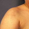
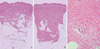
A 2-yr-old girl was presented with erythematous atrophic lesion on the right shoulder, which appeared since birth. The size of lesion was slowly growing but the shape, color, or texture of the lesion had not changed since first being discovered. Her family and medical history was unremarkable. On physical examination, she had slightly depressed erythematous plaques on the right shoulder (Fig. 1). There was no induration on the lesion. Punch biopsy for microscopic examination was done from the lesional and adjacent normal-appearing skin for comparison. Histopathological examination of a specimen from the involved skin showed markedly reduced dermal thickness when compared with adjacent normal skin (Fig. 2A, B). The collagen bundles in the dermis were slightly homogenized in appearance (Fig. 2C). The elastic tissue stain showed no abnormality.
DISCUSSION
IAPP is a form of dermal atrophy, which mostly affects women during their adolescence and young adulthood (2). In one report, of the 40 cases 26 were women and 14 were men, giving a female/male ratio of 2:1. The mean age of patients was 30 yr, although patients have been as young as 7 yr and as old as 66 yr at initial presentation. And 8 of the 40 cases developed before the age of 13 yr (3). However, none have been reported to occur with congenital lesion like our patient. The lesions are single or multiple, usually round or ovoid in shape. The affected skin is usually brown or blue to violet in color and the surface of the skin is normal in appearance with a lack of induration (4). It usually occurs on the trunk, especially on the back and lumbosacral region. Other frequently involved sites are the chest, arms, and abdomen (5). It occurs usually bilaterally and symmetrically in distribution (6). However, unilateral involvement like our case has been reported (7).
Different opinions have been suggested whether IAPP is a variant of morphea or a separate distinct entity. Recently IAPP has been regarded as a superficial form of localized morphea. It is favored by similar clinical and histological features. Lesions of "burn-out" morphea may be atrophic similar to that of IAPP (8) and some histologic similarities such as mild sclerosis and collagen homogenization exist in a few cases (1, 9). There was a case report of a patient with typical IAPP who developed progressive systemic sclerosis (10). However, whether this disorder is an end stage of morphea is unclear. Some clinicians regard IAPP as a distinct entity on the basis of the following reasons. IAPP does not show the typical lilac ring of morphea and IAPP progresses for a longer duration than morphea (1, 8). In addition, the lesions of IAPP are characterized by atrophy without any indurations, whereas morphea begins with indurations and ultimately results in atrophy (1, 11). Moreover, histological findings such as dermal atrophy and usual absence of dermal sclerosis of IAPP are different from those of morphea (1). A report that analyzed glycosaminoglycans in IAPP and morphea showed decreased amount of glycosaminoglycans in IAPP but increased amount in morphea, which suggests that glycosaminoglycans metabolism in IAPP may be unique and quite different from that in morphea (12). From this point of view, our patient could be diagnosed as IAPP. She had atrophic lesions without indurations. And the histological findings showed a significant decrease in dermal thickness when compared with adjacent normal skin, which were consistent with the findings of IAPP.
In summary, IAPP can be presented with a congenital onset and thus should be considered in the differential diagnosis of congenital skin lesions.
 XML Download