

 PDF
PDF ePub
ePub Citation
 Print
Print


INTRODUCTION
Schwannoma is a benign nerve sheath tumor that most commonly occurs solitary in otherwise normal individuals. Occasionally it presents with multiple form involving several areas along the peripheral nervous system, including cranial nerves, spinal nerve roots, the brachial and lumbosacral plexuses, or major peripheral nerves (1, 2). Patients can be definitely diagnosed with schwannomatosis if they have had two or more pathologically proven schwannomas and without radiographic evidence of a vestibular nerve tumor at age greater than 18 yr (3). If radiologic examination such as brain magnetic resonance imaging (MRI) is not available, then a probable diagnosis may be made if the patient has two or more pathologically proven schwannomas and no clinical symptoms of eighth nerve dysfunction at age greater than 30 yr or two or more schwannomas in an anatomically limited distribution without clinical finding of eighth nerve dysfunction at any age (3).
CASE REPORT
A 21-yr-old woman was hospitalized for evaluation of an incidentally found mediastinal mass on routine check. Physical examination revealed palpable soft tissue masses on the right posterior neck and right ankle. Those soft tissue masses had been found about four years previously and tumor aspiration had revealed findings of benign soft tissue tumor. She had no stigmata of NF-1 or NF-2, or other tumors. The family history was negative for neurofibromatosis. Her chromosomal analysis showed normal structural variation of chromosome 9 (44,XX,inv(9)(p11q12)) without evidence of chromosomal abnormality.
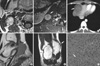
MRI of the brain revealed no intracranial tumors including vestibular schwannoma. There was a well-marginated mass in the right carotid space (Fig. 1A) and another smaller one between the right quadratus lumborum and the psoas muscle at the level of the second lumbar vertebra (Fig. 1B). Contrast-enhanced chest CT scan showed well-defined soft tissue mass in subcutaneous tissue of the right posterior neck and also an enhancing mass in the mediastinum right lateral to the heart (Fig. 1C). MRI of the ankle showed an enhancing mass below the right medial malleolus (Fig. 1D, E).
Excisional biopsy for the soft tissue masses in right carotid space and right ankle was done and en bloc resection of the mediastinal mass was performed. All the round to ovoid tumors were located within the nerves and invested with epineurial tissue. All surgically removed tumor specimens proved to be schwannomas with areas of Antoni A and B (Fig. 1F). Immunohistochemical staining revealed that most tumor cells reacted strongly for S-100 protein.
DISCUSSION
The term schwannomatosis or neurilemmomatosis has been used to describe patients with multiple nonvestibular schwannomas with no other stigmatas of NF-2 (6). Bilateral vestibular schwannomas are the classic hallmark of NF-2, with multiple schwannomas on cranial, spinal, and peripheral nerves and intracranial and intraspinal meningiomas and intramedullary ependymomas (7).
Several recent reports have suggested that some patients may develop multiple schwannomas without any associated NF-1 or NF-2 stigma (8). Such a presentation has been termed schwannomatosis or neurilemmomatosis (4, 5). Segmental forms of the entity may occur. An exact delineation between schwannomatosis and NF-2 has not been made, and an identical genetic background has been suggested. Honda et al. (9) indicated that germline mutations in the NF-2 gene were the molecular mechanism of schwannomatosis, which in this sense would represent an incomplete form of NF-2. MacCollin et al. (7) considered schwannomatosis to be a distinct clinical entity separate form NF-2. Many of previous reports described patients with highly localized disease of the peripheral nerve. Some case reports of patients with multiple schwannomas without clear anatomic localization were reported before the National Institute of Health consensus statement and include patients who would now be classified as having NF-2 (10). Patients with highly localized disease may harbor so-called "segmental" mutation of the NF-2 gene or other schwannoma-related genes (7). Alternatively, they may have suffered a single early transforming event with local noncontiguous spread of tumors such as that postulated for patients with multiple meningiomas without family history. Those persons with more generalized disease may carry a tumor suppressor gene syndrome, as their phenotype is similar to more common tumor suppressor syndromes (11). Multiple schwannomas in a child is a difficult clinical problem because the appearance of peripheral schwannoma may precede that of vestibular schwannoma in patients known to have NF-2 (7).
Some differences have become apparent between schwannomatosis and neurofibromatosis with respect to their clinical manifestations. First, the patient age at presentation differs. Schwannomatosis is presented at middle age. This age corresponds to that for most patients with sporadic schwannomas, but contrasts with the earlier age of less than 20 yr for patients with NF-2. As for our patient, she was detected without symptom incidentally after 20 yr of age. The second difference with neurofibromatosis is the absence of any family history in schwannomatosis. Additionally, the genetic mechanism of schwannomatosis differs from that of NF-2 (12). There is no evidence of germline mutations on direct sequence of the NF-2 gene among schwannomatosis patients.
Radiologic features of tumors in schwannomatosis are the same with those of schwannoma, except multiplicity on different anatomical locations. Schwannomas are iso- or slightly hypodense on non-enhanced CT scans. Small tumors usually show uniform enhancement, while larger lesions may have a heterogeneous pattern. On MR imaging, schwannomas are slightly hypointense or isointense on T1-weighted image, and show increased signal intensity on T2-weighted image. Most schwannomas show intense homogeneous enhancement following contrast administration.
To summarize, of patients presenting with schwannoma, 3 to 4% are found to have multiple lesions (8). The diagnosis of schwannomatosis seems justified for adult patients presenting with two or more schwannomas in different anatomical locations who show no clinical or radiological signs of a vestibular nerve tumor and no meningiomas, ependymomas, or any other signs of NF-2. We demonstrate that this rare case of schwannomatosis in a 21-yr-old woman with no associated with NF-1 or NF-2, discontinuously involving peripheral nerves of the right side body.
 XML Download