

 PDF
PDF ePub
ePub Citation
 Print
Print


INTRODUCTION
Coronary artery fistula is not a rare anomaly characterized by abnormal connections between coronary arteries and cardiac chambers or pulmonary vasculatures. It is usually congenital (1, 2), resulting from incomplete obliteration of the primitive myocardial sinusoids and fistulous tracts. On the other hand, acquired coronary artery fistulas are extremely rare and have been reported as complications after chest trauma (3), myocardial infarction (4), myocardial biopsy (5), and cardiac surgery (6-12). Iatrogenic fistulas between the coronary artery and the left ventricle after septal myectomy for hypertrophic obstructive cardiomyopathy (HOCM) have been also reported sporadically and scarcely (6, 7, 12-14). Therefore, its proper management principles and prognosis are not well known. We report one case of spontaneous closure of an iatrogenic coronary artery fistula draining into the left ventricle that occurred after surgical septal myectomy in a patient with HOCM.
CASE REPORT
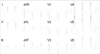
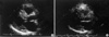
A 46-yr-old woman was hospitalized for evaluation of chest pain and shortness of breath that began three months ago. She had recurrent episodes of anterior chest pain, which was relieved by sublingual nitroglycerin. On physical examination, her blood pressure was 130/70 mmHg, pulse rate 54 beats per minute, respiratory rate 20 per minute, and body temperature 36℃. Jugular venous engorgement was not found. A grade III systolic murmur was audible on her left lower sternal border. The lung sounds were clear. Mild pretibial pitting edema was found in both lower extremities. Electrocardiogram revealed regular sinus rhythm and left ventricular hypertrophy with strain pattern (Fig. 1). Chest radiograph showed moderate cardiomegaly without pulmonary edema (Fig. 2). On peripheral blood test, WBC count was 7,590/µL, hemoglobin 12.0 g/dL, platelet count 291,000/µL, serum total protein 6.2 g/dL, albumin 4.1 g/dL, and N-terminal proBNP 3,800 pg/mL. Arterial blood gas analysis and other biochemical tests were normal. Preoperative coronary angiography revealed no stenosis or abnormal connections of the coronary arteries. Transthoracic echocardiography demonstrated diffuse concentric hypertrophy of the ventricular myocardium, which was predominant in the septum (Fig. 3). The septal wall thickness was 31 mm and the posterior wall thickness was 12 mm, and the ejection fraction was normal. No regional wall motion abnormalities were found. Systolic anterior motion of the mitral valve leaflets (SAM) with moderate amount of mitral regurgitation and mid-systolic closure of the aortic valve were observed. Resting peak pressure gradient in the left ventricular outflow tract (LVOT) was 71 mmHg, and it increased up to 114 mmHg with Valsalva maneuver. Left atrial enlargement and moderate pulmonary hypertension were also noted.
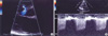
With these findings, the patient underwent surgical septal myectomy, and myocardial tissue was resected from the basal part of the septum (Fig. 3). Three days after the surgery, postoperative echocardiography showed a reduction of septal wall thickness to 9 mm, no SAM, and the peak pressure gradient of the LVOT was only 16 mmHg. However, color Doppler imaging revealed a diastolic blood flow originating from the interventricular septum directed into the left ventricular cavity, i.e. iatrogenic coronary artery fistula to the left ventricle (Fig. 4). Ten days after the surgery, the patient complained of mild chest discomfort, which was relieved by sublingual nitroglycerin. Diagnostic coronary angiography was done, but no fistulas from septal branches of the left anterior descending coronary artery artery to the left ventricular cavity could be found. Follow-up transthoracic echocardiography showed no residual shunt flow that had been present seven days before. We concluded that the iatrogenic coronary artery fistula closed spontaneously.
DISCUSSION
In the current report, we describe an acquired coronary artery fistula to the left ventricular cavity after surgical myectomy for the treatment of HOCM. The fistula appeared at the site of surgical intervention and involved septal perforator branches that had been cut surgically, thus creating a communication between the branches and the LV outflow tract. It resulted in a left-to-left shunt between septal perforators of the left anterior descending coronary artery and the left ventricle. There have been sporadic case reports of acquired coronary artery fistulas to the left ventricle after a septal myectomy for HOCM (6, 12, 13). Chenzbraun et al. (7) reviewed postoperative echo-Doppler studies of 26 consecutive patients who had undergone myectomy for HOCM between 1988 and 1991 and found coronary artery fistulas in 19%. They concluded that fistulas to the left ventricle after myectomy might occur more frequently than expected, but might be easily missed on echocardiographic examination unless specifically sought.
If the shunt amount of the fistula is clinically significant, the distal coronary circulation may be at risk for coronary steal and myocardial ischemia (15). Meanwhile, smaller fistulas may impose minimal or no hemodynamic burden. However, persistent fistulas may expose the patient at risk for infective endocarditis, congestive heart failure, ischemia, accelerated atherosclerosis, or rarely, rupture (16-18).
In our case, the fistula was detected on routine postoperative transthoracic echocardiographic examination. Color Doppler scanning of the intramyocardial coronary artery at basal septum showed a continuous turbulent flow connecting to the left ventricular cavity. This suggests that echocardiographic examination is useful for detection of acquired coronary fistulas (19) and may be used for noninvasive follow-up of these abnormalities. Other imaging modalities such as magnetic resonance imaging, computed tomography, and radionucleotide cine-angiography have been reported to aid in the diagnosis (20, 21) and act as an assisting tool for coronary angiography, which can directly visualize a fistulous connection (22).
The management scheme and the natural history of acquired coronary fistulas to the left ventricle have not been determined due to their low incidence (10). Surgical treatment has been favored for the management of fistulas developed after chest trauma and for those that drain into right-sided chambers which tend to become worse progressively because of the high-to-low pressure communication (3). However, previous reports for the fistulas that drain into the left ventricle such as the current case suggest that medical therapy appears to be an appropriate treatment strategy (6, 13). This is also supported by an observational report of a coronary artery fistula that remained stable thirty years after myectomy for septal hypertrophy (12).
In conclusion, acquired coronary artery fistula to the left ventricle can occur after surgical septal myectomy, and transthoracic Doppler echocardiographic examination is a useful method for its diagnosis and follow-up. Although its treatment principle is not determined yet, conservative management seems to be sufficient because its clinical course is likely to be benign.

 XML Download