

 PDF
PDF ePub
ePub Citation
 Print
Print


INTRODUCTION
Benign stenosis involving the main bronchi is ideally suited for a bronchoplastic procedure because this requires only minimal clear margins for a cure and it preserves lung parenchyma. However, restrictions have been recommended for bronchoplasty because of concerns about fatal postoperative complications occurring in otherwise non-lethal diseases. Instead, alternatives to surgery such as laser, balloon dilatation, or stent insertion have been used as mainstays of therapy. However, the facts that there is no ideal stent available to use for permanent placement without complications and that the removal or repositioning of a metal stent is sometimes possible (1) but technically demanding shows such alternative modalities cannot be proper solutions to benign bronchial stenosis. Yet in recent years, there have been several reports on the successful results of bronchoplasty with low operative mortality and morbidity (2, 3). The operative mortality rate of recent studies was less than 5% and anastomosis-related problems were uncommon (4).
Since 1995, we have treated the patients with benign bronchial stenosis under a set of coherent treatment guidelines. In this study, we have reviewed our results of main bronchial reconstruction operation that were done over the last 10 yr and we provide the validity and proper clinical information on using bronchoplasty for benign main bronchial stenosis.
MATERIALS AND METHODS
Patients
From 1995 to 2004, 28 consecutive patients underwent surgical treatment, including mainstem bronchoplasty with sparing of pulmonary parenchyma, for benign bronchial stenosis at the Samsung Medical Center. Data were collected by reviewing the hospital and office records. Twenty-one patients were female and 7 were male. The mean patient age was 34.4 yr, and their ages ranged from 2 to 69 yr.
Preoperative conditions
Most of the patients (25 out of 28) had respiratory symptoms at the time of diagnosis such as cough, dyspnea, sputum, wheezing, hemoptysis with or without fever. In three asymptomatic cases, the diagnostic workup was started from the unresolved atelectasis on chest radiograph which was incidentally found for other reasons. All of the enrolled patients underwent fiberoptic bronchoscopy, a standard chest radiograph and thoracic CT. On the standard radiograph, lobar or segmental atelectasis (n=18), pneumonia (n=2), pulmonary fibrosis, bronchiectasis, a mass shadow in the main bronchus were identified. Two patients did not show any abnormality on standard chest radiograph. Preoperative pulmonary function tests were available for 20 patients, and the tests showed that the average forced expiratory volume for 1 sec (FEV1) was 1.76 L (64% of the normal predicted value), with a range from 0.89 L to 3.57 L (29% to 114% of the normal predicted value).
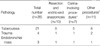
All the patients underwent main bronchus reconstruction for stenosis caused by benign diseases including tuberculosis (n=21), trauma (n=4), or benign endobonchial mass (1 endobronchial leiomyoma, 1 inflammatory pseudotumor and 1 lipoma). In all of the cases of traumatic main bronchial stenosis, diagnosis was not suspected until the atelectasis was not resolved by more than 2-weeks of pulmonary toileting. Endobronchial masses were pathologically confirmed before the operation with bronchoscopic biopsy. Twenty of all tuberculous main bronchial stenosis patients had histories of anti-tuberculosis medication. None of the patients arbitrarily stopped their medication and the duration of the medication ranged from 6 months to 12 months. One of the tuberculous stenosis patient started anti-tuberculosis medication after initial bronchoscopy due to actively caseating and stenotic endobronchial tuberculosis and then surgically treated after 3 months of medication. The average duration from the completion of medication to the time of operation was 116 months, with a range from 6 months to 468 months.
For the patients showing pneumonia or atelectasis on their preoperative radiograph, aggressive toileting of sputum (including therapeutic bronchoscopy) and two weeks antibiotic coverage before operation were applied.
Preoperative non-surgical intervention and operative indications
Nine patients who had fibrous stricture due to tuberculosis (n=8) or trauma (n=1) underwent intervention treatment before operation; balloon dilatation under the rigid bronchoscope was performed in three patients; removal of granulation tissue under rigid bronchoscopy was done in one patient and application of stent was done in five patients. All of them showed no improvement with the non-surgical intervention.
For the patients showing main bronchial stenosis we initially performed a thorough examination with flexible bronchoscopy and a CT scan. If anatomical correction was necessary, we then tried rigid bronchoscopic evaluation and we tried non-surgical interventions such as laser, balloon dilatation, or stent insertion, as well as controlling the underlying infectious disease. However, if surgical treatment was determined to be helpful to the patients or failure of non-surgical intervention was determined, we then performed bronchoplastic procedures without any unnecessary delay. When the patients were less than 40 yr, they had adequate pulmonary function tolerating general anesthesia and thoracotomy, and lung parenchymal destruction accompanied by bronchial stenosis was none or minimal, we enthusiastically recommended bronchoplastic operation.
Operative techniques
Single or double-lumen endotracheal tubes were used for the safe conduct of anesthesia. Posterolateral thoracotomy of the involved bronchial side was usually used, however, when carinal reconstruction was intended, right posterolateral thoracotomy was used. Dissection and mobilization of the distal trachea and the bilateral main bronchus was performed in most of the cases. In every case, end-to-end anastomosis was done using 4-0 or 5-0 Vicryl (Ethicon, Somerville, NJ, U.S.A.). Interrupted sutures were always used and placed starting at both ends of the resection; they were evenly spaced and were added one by one from each side to reach the side opposite to the small part of the bronchus and were tied after all the sutures had been placed. Simple resection and end-to-end anastomosis between the distal trachea and distal main bronchus was sufficient in these cases. However, in 19 patients, concomitant procedures such as lobectomy, wedge resection of lung, or carina reconstruction were necessary. The indications and methods of the concomitant procedures are listed in Table 1. In order to release the tension around the anastomosis, mobilization of pulmonary ligament was sufficient in 14 cases. But, additional mobilization techniques were needed in 14 patients: pretracheal release for 4, hilar release for 1, and cervical flexion for 1. Especially for left side main bronchoplasty, mobilization of the aortic arch by detaching the ligament of Botalli and Marshall's fold (n=8) was needed. The anastomosis was wrapped with pedicled pericardial fat (n=1) or parietal pleura (n=3). At the end of the operation, before extubation, the bronchial tree was inspected using flexible bronchoscope to evaluate the suture line, the patency, and the alignment of the bronchial anastomosis and to aspirate secretions and blood.
Follow-up bronchoscopy after the operations was done on postoperative day seven and at approximately 2 months. Chest CT with volume rendering reconstruction of the airway was also done at postoperative 2-month. If the bronchoscopy results were good, only the chest CT was checked at 6 month, and then it was checked every year.
RESULTS
The mean hospital stay was 14.7 days (range: 8 to 38 days). There was no operative mortality and no postoperative complications requiring re-operation. There were 2 postoperative morbidities. Two patients had retained secretions, and this was resolved with bronchoscopic toileting or endotracheal intubation for less than 5 days. There were minor complications such as persistent air leak and persistent chest tube drainage in seven cases, but these were not related to main bronchial anastomosis site based on the follow-up bronchoscopic finding. There was no patient who had recurrence of the airway stenosis, although two patients had mild growth of the granulation tissues, as was seen by follow-up bronchoscopy.
Postoperative pulmonary function data were available for 12 patients, and all of these patients showed an increase in the FEV1 with a range of 6% to 32% (mean increase of FEV1: 0.5 L). Even for the patients who did not perform follow-up pulmonary function test, no patient showed limited daily activity or work activity. The mean follow-up time was 19.6 months (range: 0.5 to 91 months). All of the patients are still alive without symptomatic obstructive airway problems.
DISCUSSION
Since Clement Price Thomas first performed mainstem bronchial sleeve resection in 1947 (5), bronchoplasty has been the choice of treatment for the patients having central or main bronchial tumors with poor pulmonary function, which is a medical condition not suitable for pneumonectomy. However, the extended use of bronchoplasty procedure even for benign main bronchial stenosis is still controversial because this disease has been considered as not being fatal, but rather, it is an uncomfortable malady. Furthermore, because various postoperative complications such as empyema, hemothorax, arrhythmia, atelectasis, pneumonia, prolonged air leak, vascular erosion, and vocal cord paralysis have been reported (6), bronchoplasty had not been recommended as the first choice of treatment for benign main bronchial stenosis.
In recent years, several reports have suggested that metal stent should be a good alternative to surgery for bronchial stenosis (7, 8) because 1) the metal stent can be inserted through an endotracheal tube under local anesthesia (9), 2) it does not impair the drainage of sputum because ciliary movement is not interrupted (10), and 3) if it covers another non-stenosed bronchus, lung ventilation will not be blocked because of the spaces between the wires. However, the metal stent may not be suitable, especially for benign bronchial stenosis, because of such possible serious complications as fatal hemorrhage, fistula formation, migration, and embedding in the bronchial mucosa. In addition, any recklessly forced attempt at relocation or removal of a stent may be lethal because of encroachment on the adjacent blood vessels by the spike of stent (11).
Recent reports suggest that covered self-expandable metallic stents for benign airway disorders can be removed (1). But most of the indications for removal were complication of the stent itself such as excessive granuloma formation, stent failure, or stent fracture. Only 2 of the ten removal cases were accomplishment of treatment. In the report removal rate was 25.6% and this rate was higher than our operative complication rate.
In fact, bronchoplasty is not considered risky any more (12). In the current study, there was not any mortality, serious complication or recurrence of the airway problems. In the series of 79 patients who underwent bronchoplasty or angiobronchoplasty for malignant endobronchial neoplasm (4), the authors asserted that bronchoplasty was a safe procedure. The operative mortality rate of the study was 5.1% and only 3 of the enrolled patients had anastomosis-related problems; there were 2 cases of dehiscence and 1 case of stenosis. Another study on 22 bronchoplasty patients with malignant or benign bronchial obstruction (2) emphasized that bronchoplasty can be safely performed; there were only 3 postoperative complications and no operative mortality. From our personal experience in our hospital, we had 150 patients with bronchial sleeve lung resection for lung cancer with 2 percent mortality and only 1 case of an anastomotic problem (unpublished data).
However, bronchoplasty for the benign disease is technically a little different from that for the malignant disease. Because there is no need to assure a sufficient resection margin, it is easier to get a tension-free anastomosis without performing any releasing procedures. Yet because an inflammatory process or fibrosis involved long segments of the bronchi, the airway for the anastomosis was generally very unhealthy and, therefore, the necessary careful technique by the surgeon is very demanding.
The promising results of our study may be attributed, to some extent, to the preoperative preparation and the postoperative care. We recommend at least 3-months of anti-tuberculosis medication and preferably a negative conversion of tuberculosis bacilli on the sputum smear before performing operations for the patients having history of tuberculosis. The routine postoperative intensive hemodynamic and respiratory care, close monitoring, and early ambulation also contributed to the uncomplicated recovery.
Tuberculous stenosis patients consist mainly of our patients group and this is one of the most prominent characteristics. The possible cause is that Korea is one of the endemic areas of tuberculosis for the past several decades (13). Lee and Chung (14) classified endobronchial tuberculosis lesions into 7 subtypes by their own criteria and contended that some specific types of endobronchial lesions changed into fibrostenotic type during anti-tuberculosis medication. In addition, they observed that fibrostenotic tuberculous lesions did not improve despite anti-tuberculosis chemotherapy. These features of endobronchial tuebrculous lesions can be the cause of tuberculous main bronchial stenosis.
Our study included 8 carina-involving operations. Carina resection and reconstruction is still a technically challenging procedure to surgeons, and the potential for morbidity and mortality is still quite high. Mitchell and co-workers (15) reported that patients undergoing carinal and lobar resection had anastomotic morbidity as high as for carinal pneumonectomy. Our results were without mortality and significant mortality, and this can be explained by the above-mentioned protocol of pre- and postoperative management and that our operative techniques focused on the principles of tension-free anastomosis and avoidance of excessive dissection for preserving the blood supply.
In conclusion, mainstem bronchoplasty with sparing of the pulmonary parenchyma for the patients with benign main bronchial stenosis can be a safe procedure without operative mortality and serious morbidities. In the report by Nonoyama et al. (16), mainstem bronchial resection has been shown to restore both airway patency and lung function after atelectasis conditions lasting up to 9 yr. Therefore, if the procedure is anatomically feasible, mainstem bronchoplasty should be considered as one of the primary treatment modalities.
 XML Download