

 PDF
PDF ePub
ePub Citation
 Print
Print


INTRODUCTION
CD99 is a 32-kDa cell surface sialoglycoprotein, which is expressed in a wide variety of human cells. Functionally, CD99 is considered to act as an adhesion molecule or as a signal-transducing molecule (1, 2). Since original report that demonstrated its diagnostic utility in Ewing's sarcoma/primitive neuroectodermal tumor (3), CD99 expression has been demonstrated in a broad range of normal and neoplastic cells (4-10). In hematologic neoplasms, CD99 expression is well correlated with TdT positivity (11) and has been observed in T- and B-lymphoblastic lymphomas/leukemias, 55% of chloromas, 43% of acute myelogenous leukemias, and chronic myelogenous leukemias in blastic crisis (12). While CD99 is considered a useful marker for detecting lymphoblastic neoplasms, previous studies on mature T- and B cell non-Hodgkin's lymphomas (NHLs) have reported discordant results concerning the spectrum of CD99-positivity (11, 13, 14). In this study we examined CD99 immunoreactivity in a variety of NHL, including 21 lymphoblastic lymphomas/leukemias, 87 mature B-cell lymphomas, and 74 mature T and NK cell lymphomas.
MATERIALS AND METHODS
Case selection
During the period from January 1996 to April 2004, 182 patients diagnosed as having non-Hodgkin's lymphoma were randomly selected at Samsung Medical Center, Seoul, Korea. The selected cases comprised 21 lymphoblastic lymphomas/leukemias, 37 diffuse large B cell lymphomas, 18 Burkitt's lymphomas, 12 follicular lymphomas, 11 small lymphocytic lymphomas, 9 mantle cell lymphomas, 28 NK/T-cell lymphomas, 8 angioimmunoblastic T-cell lymphomas, 23 peripheral T-cell lymphomas, and 15 anaplastic large cell lymphomas. Clinical and laboratory records including a chromosomal study were reviewed.
Histologic examination
Hematoxylin eosin-stained slides were reviewed in all cases and the diagnosis was made based on recent WHO classification (15).
Immunohistochemical studies
Paraffin sections were stained with monoclonal CD99 antibody, 12E7 (1:40, DAKO, Carpinteria, CA, U.S.A.) in an automated staining system. Antigen retrieval was conducted in a microwave oven in citrate buffer at pH 6.0 before incubation with primary antibody. The tumors were considered immunoreactive for CD99 if diffuse cell membrane staining was observed in more than 70% of tumor cells. Focal or weak staining was interpreted as negative. In addition, paraffin sections were stained with ALK1 monoclonal antibody at a dilution of 1:40 (DAKO, Glostrup, Denmark), polyclonal antibody for CD3 (1:200, Novocastra, U.K.), monoclonal antibody for CD20 (1:500, Novocastra), monoclonal antibody for bcl-2 (1:40, DAKO), polyclonal antibody for bcl-6 (1:40, Santacruz, California, U.S.A.), monoclonal antibody for CD10 (1:50, Novocastra, U.K.), monoclonal antibody for CD56 (1:20, Monosan, New Zealand), monoclonal antibody for CD30 (1:30, DAKO, Glostrup, Denmark), and with monoclonal antibody for CD21 (1:25, DAKO, Glostrup, Denmark).
Western blot analysis to verify CD99 expression
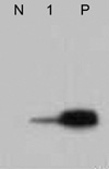
To verify CD99 expression in anaplastic large cell lymphoma, western blot analysis was performed in one case of anaplastic large cell lymphoma of which fresh tumor tissue was available. For the positive control, lymphoblastic lymphoma was used. Monoclonal CD99 antibody, 12E7 (DAKO, Carpinteria, CA) was applied at a dilution of 1:1,000.
RESULTS
The results of immunohistochemical staining for CD99 are summarized in Table 1.
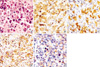
CD99 was positive in 100% of T-lymphoblastic lymphomas, but in only 60% of B-lymphoblastic lymphomas/leukemias. The majority of mature T and NK cell lymphomas were negative for CD99, except anaplastic large cell lymphomas. Fifty-four percent (8/15) of systemic ALCLs reacted with anti CD99 antibody. ALK positive tumors expressed CD99 more frequently than ALK negative tumors; 7 of 10 (70%) ALK positive ALCLs expressed CD99 (Fig. 1), whereas only 1 of 5 (20%) ALK negative ALCLs were positive. The intensity of CD99 in ALCL was rather weaker than that of lymphoblastic lymphoma, but cytoplasmic membrane stain of tumor cells was obvious which was in accord with the result of western blot analysis (Fig. 2). Of the mature B cell lymphomas, 5.3% (2/37) of diffuse large B cell lymphomas and 11.1% (2/18) of Burkitt's lymphomas were positive for CD 99. All follicular lymphomas, small lymphocytic lymphomas, and mantle cell lymphomas were negative for CD99.
Of the 2 cases of CD99-positive diffuse large B cell lymphoma, one was a 51-yr-old man who presented with axillary lymph node enlargement. This tumor was composed of large cells, which were positive for CD20, bcl-6, MUM-1, and EBV ISH, and negative for CD3 and CD10. The second patient was a 56-yr-old female who presented with an enlarged neck lymph node; tumor cells were large, had a diffuse appearance, and were positive for CD20 and bcl-6, and negative for CD10, bcl-2, MUM-1, and EBV ISH.
In the two CD99-positive cases of Burkitt's lymphomas, one patient was a 78-yr-old man who present with a stomach lesion. Tumor cells showed strong bcl-6 and CD20 expression, but were negative for bcl-2, and had a Ki-67 labeling index more than 99%. The other patient was 65-yr-old man with a retroperitoneal tumor, which was negative for TdT and positive for CD79a and CD20 with a Ki-67 labeling index of 90%. In this case bone marrow metastasis was identified at the initial diagnosis and a chromosomal study revealed t(8:14). Microscopically tumor cells were monotonous with round to oval nuclei and a prominent nuclear membrane. Nucleoli were prominent and ranged from 2 to 5 per nucleus. Occasional tumor cells had a centrally located single prominent nucleolus resembling an immunoblast. The chromatin was coarse and irregularly distributed.
DISCUSSION
CD99 is a 32-kDa transmembrane sialoglycoprotein that is expressed on many types of human cells. However, despite its broad distribution CD99 is only strongly expressed in relatively a few cell types, such as bone marrow progenitor cells, pancreatic islet cells, granulosa cells of the ovary, and Leydig and Sertoli cells of the testis and ependyma (3, 7, 16). CD99 was originally described as a diagnostically useful marker for Ewing's sarcoma/primitive neuroectodermal tumor (3), but CD99 immunoreactivity has also been documented in a variety of other tumors, including sex cord tumors of the testis and ovary, synovial sarcoma, chondrosarcoma, and neuroendocrine tumors (6-8). Moreover, within the hematopoietic system CD99 expression is strongly correlated with TdT expression, in addition it is highly expressed in early CD34+ precursors, which is in line with the maturation process (16, 17). In hematologic neoplasms, CD99 expression has been observed in T-and B-lymphoblastic lymphomas/leukemias, chloroma, acute myelogeneous leukemia, and chronic myelogeneous leukemia in blastic crisis (12, 13).
The expression of CD99 in non-Hodgkin's lymphomas, other than those of the lymphoblastic type has become the subject of debate. One study had reported a negative reaction in all NHLs, other than lymphoblastic lymphoma (9), whilst the other had described only weak, non-uniform, poorly localized cytoplasmic staining in a minority of cases (13). Vartanian et al. (14), using a heat-induced epitope retrieval (HIER) technique, reported that a variety of malignant lymphomas, other than the lymphoblastic type, show significant CD99 reactivity. However, Robertson et al. (11), also using HIER, found that positive CD99 staining was restricted to lymphoblastic disorders, and ascribed this discrepancy to technical differences. The present study, also using HIER, revealed distinct membranous CD99 staining in a few cases of NHL of the mature phenotype, and unexpectedly high expression in ALK-positive ALCLs. Twenty percent of ALK-negative ALCLs expressed CD99, and this rose to 70% in ALK-positive ALCLs.
How the high expression of CD99 in ALK positive ALCLs be explained? Though there appears to be no clear-cut answer to this question, present data suggest that ALK protein may play a role. CD30 is a member of the tumor necrosis factor receptor (TNFR) superfamily and one of essential findings for the diagnosis of ALCL and Hodgkin's disease (18, 19). In Hodgkin's disease (HD), constitutively activated NF-κB by Hodgkin's disease (HD), constitutively activated NF-κB by either EBV LMP-1 gene or CD30, down-regulates the expression of CD99 and the repression is restored by the inhibition of NF-κB activity (20). In ALK-positive ALCL, NF-κB activation is not observed in the presence of equivalent levels of CD30 expression (21) because NPM-ALK oncoprotein abrogates CD30 signaling and constitutive NF-κB activation (22).
The functional roles of CD99 expressed in ALCL are unknown. The CD99 signal enhances Fas-mediated apoptosis in the human leukemic cell line (23). Engagement of distinct epitopes on CD99 rapidly induces T cell death by a novel caspase-independent pathway through reorganization of cytoskeleton (24). Because ALK-positive ALCLs show higher apoptosis rate and higher levels of active procaspase 3 and proapoptotic Bcl-2 family proteins such as BAX and BCL-XS than ALK-negative ALCLs, it is conceivable that CD99 works on apoptotic cell death in ALK-positive ALCLs (25, 26).
In summary, present study demonstrates that CD99 expressed frequently in ALK-positive ALCL, but not in other mature T and NK cell lymphoma. The result expands our understanding on the spectrum of CD99-positive neoplasm although its biologic and clinical significance remained to be clarified.
 XML Download