INTRODUCTION
Lymphangiomyomatosis (LAM) is a rare disease of a hamartomatous nature characterized by proliferation of abnormal lymphatic smooth muscle cells. Smooth muscle cells are often classified in the family of perivascular epithelioid cells (PEC). Some of these cells include renal angiomyolipoma, sugar tumor of the lung and pancreas, and clear-cell myomelanocytic tumor of the falciform ligament. These so-called "PECom" all express HMB-45 (1). The primary site of origin is the lung and occurrence is usually associated with decreased pulmonary function and chylous effusions (2). The outcome of this disease can be devastating but the underlying etiology and effective treatments are unknown. Extrapulmonary LAM is quite rare and mainly located in the pelvis and retroperitoneum, along the lymphatic channels. The clinical features are the presence of chylous ascites, a palpable abdominal mass, and abdominal pain (3). We report a case of a young female who was initially diagnosed with retroperitoneal LAM that eventually led to pulmonary involvement with decreased lung function.
CASE REPORT
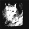
A 21-yr-old female patient presented with a sudden onset of low abdominal pain. With the clinical impression of an ovarian cyst, she was transferred from a local GYN clinic to our hospital. Gynecologic ultrasonography revealed a large posterior pelvic mass with an irregular echogenicity measuring 9.7×4.2 cm in size (Fig. 1). The patient had no specific past medical history with a parity of 0-0-0-0. A considerable amount of fluid was present in the cul de sac. The uterus and both adnexae were unremarkable. There was no ultrasonographic evidence of lymphadenopathy. Pelviscopy showed a large, thin walled, partly cystic, pelvic mass measuring approximately 10 cm in diameter. The mass was adherent to the internal and external iliac vessels and the right pelvic wall. It was not possible to remove the tumor completely. The retroperitoneal mass was partly removed by pelviscopy and samples were submitted for frozen section analysis.
Pathologic findings
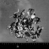
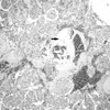
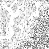
The submitted specimen for frozen section analysis consisted of multiple irregular fragments of soft tissue weighing approximately 30 g. The tumor was soft and grayish-tan with focal areas of hemorrhage (Fig. 2). On frozen section, the possibility of epithelioid smooth muscle tumor or paraganglioma was suspected. Microscopically, the masses were characterized by a haphazard proliferation of smooth muscle cells (LAM cells) arranged in fascicular, trabecular, and papillary patterns around a ramifying network of endothelium-lined spaces. The cells were plump or epithelioid with abundant eosinophilic cytoplasm and bland nuclei with no mitotic activity. These cells invaded into fat tissue surrounding the lesion. The vascular spaces were empty or sometimes filled with eosinophilic material (Fig. 3). Aggregations of lymphocytes were between the LAM cells, representing vestiges of preexisting lymph nodes (Fig. 4). Occasional foci of hemorrhage and necrosis were present.
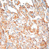
Most of the LAM cells showed a positive reaction for alphasmooth muscle actin (Dako Co., Carpinteria, CA, U.S.A.), vimentin (Zymed, San Francisco, CA, U.S.A.), desmin (Bio-Genex, San Ramon, CA, U.S.A.), and HMB-45 (Dako) (Fig. 5). S-100 protein (DiNonA Inc. Seoul, Korea), cytokeratin (Dako), chromogranin (Zymed), and CD 34 (Immunotec, Marseille, Cedex, France) were negative. Estrogen (Immunotec) and progesterone (Novocastra Lab. Ltd. Newcastle, U.K.) receptors were observed in only 10% of the LAM cells. Ki-67 (Zymed) was positive in less than 5% of the tumor cells. Clinical evidence of tuberous sclerosis was not present. Evaluation for the TSC gene was not performed.
Clinical course
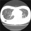
After operation, the patient had continuous oozing of chylous ascitic fluid of approximately 1,500 to 2,000 mL per day. There was no clinical or radiological evidence of pulmonary involvement at the time of the initial diagnosis. Chylous pleural effusion was also identified two months after the operation. Chest CT scans showed diffuse, small, thin walled cystic lesions in both lung parenchymas with pleural effusion (Fig. 6). The patient was treated with tamoxifen and medroxyprogesterone, but the response was limited. Even after chest tube insertion and pleurodesis, she continued to drain approximately 800 to 2,300 mL of chylous fluid per day. In the terminal stages of the disease, the patient suffered severe respiratory distress and died six months after initial operation.
DISCUSSION
Zamboni et al. (1) described a group comprising sugar tumor, angiomyolipoma, and lymphangiomyoma as a special family of tumors. The nature of perivascular proliferation of abnormal smooth muscle cells and common antigenic expression of HMB-45, a melanosomal antigen, leads to the concept of so-called perivascular epitheloid cells-PEComa.
HMB-45 positive cells are found in both LAM and angiomyolipoma; however, angiomyolipomas are characterized by the presence of abundant adipose tissue and by disorganized components of blood vessels (4). The incidence of angiomyolipomas in patients with LAM has been reported to be as high as 60% (2). Matsui et al. (3) regarded angiomyolipomas as an unusual but distinct expression of the proliferative capacity of LAM cells. Most of the tumor cells in this case showed a positive reaction for HMB-45. Accumulation of fat tissue within the lesion was not prominent in this case.
LAM is a tumor of the lymphatic channels and lymph nodes clinically manifested by recurrent chylous pleural effusions and ascites. The disease is usually progressive and unresponsive to surgery, chemotherapy, or irradiation. LAM of the lungs is characterized microscopically by abnormal smooth muscle proliferation causing gradual obstruction of small airways, lymphatics, and vasculature (5). The typical presenting symptoms include dyspnea (83%) and pneumothorax (69%) (2).
Extrapulmonary LAM is rare. Recently, Jaiswal et al. (6) reviewed 32 extrapulmonary LAM cases, including 22 cases of extrapulmonary LAM reported by Matsui et al. (3). Among the reported cases of clinically significant extrapulmonary LAM, 14 cases occurred in the lower retroperitoneum or pelvic cavity (3, 4, 6). Most extrapulmonary LAM lesions occurred in lymph nodes along the lymphatic vessels of the posterior mediastinum, retroperitoneum, and the pelvic cavity. The diagnosis of extrapulmonary LAM preceded that of pulmonary LAM in 13 of 17 patients for whom follow-up data were available (3). In this case, extrapulmonary manifestations preceded chylous pleural effusion. Our patient presented with a sudden onset of low abdominal pain. In Korea, approximately 24 cases of LAM have been reported in the current literature, all with a primary site in the lungs (7-9). This case is, to our knowledge, the first extrapulmonary LAM reported in Korea.
The origin and cause of this disease are yet to be undetermined however there are some common peculiar aspects. LAM can be divided into a sporadic form, as in this case, or a tuberous sclerosis-associated form (10-12). There have been many studies regarding the genetic predisposition of LAM, especially associated with the mutation of tuberous sclerosis genes (TSC1 and TSC2) (12, 13). This condition is usually defined as tuberous sclerosis complex (TSC). TSC is an autosomal dominant disorder and is manifested as seizures, mental retardation, autism, and hamartomatous tumors of the brain, heart, kidney, lung, and skin. These tumors include cerebral cortical tumors, subependymal giant cell astrocytomas, retinal hamartomas, cardiac rhabdomyomas, renal angiomyolipomas, and facial angiofibromas (13). Four of the 32 extrapulmonary LAM cases were associated with tuberous sclerosis (6). There was no clinical evidence of TSC in this case.
The speculation that LAM is a female sex hormone dependent tumor is supported by the high prevalence rate in women of reproductive age and exacerbation of the disease by pregnancy or by administration of estrogen. Numerous studies and case reports regarding clinical trials of hormonal therapy had been reported (14, 15). However, the role of estrogen and progesterone receptors in proliferation of smooth muscle and the therapeutic efficacy of hormone therapy are not well established. Our patient was treated with tamoxifen and medroxyprogesterone, but her response was limited.
Despite a variety of treatment regimens developed since the first description of LAM, patient survival has not improved appreciably. LAM progresses slowly and most patients die within 10 yr of diagnosis (16). Despite the slow progression rate of other LAM cases, our patient died of the disease six months after operation. The reason for her rapid progression is not clear. Prolonged chylous peritoneal and pleural effusions, aggressive deterioration of pulmonary function, and her lack of response to various treatment modalities were major contributing factor.


 PDF
PDF ePub
ePub Citation
 Print
Print


 XML Download