

 PDF
PDF ePub
ePub Citation
 Print
Print


INTRODUCTION
The rapid increase in motor vehicular traffic and its emissions have paralleled a worldwide increase in the incidence of allergic diseases such as asthma and rhinitis. A steady increment of diesel-engine-powered cars increased concentration of diesel exhaust particulates (DEP) in the atmosphere of urban areas. Because DEP contain carbon nuclei that absorbed a variety of toxic compound, including polyaromatic hydrocarbons, quinines, and aldehydes (1, 2), estimating the risk of DEP has recently become a major worldwide health issue. DEP remains buoyant in the atmosphere, with a certain fraction being carried into lungs (3). Many studies have suggested that DEP may cause lung cancer, pulmonary fibrosis, edematous change, chronic alveolitis, and bronchitis (4-6). However, few studies have investigated the relationship of DEP to bronchial asthma, which have shown increasing prevalence in urban areas of the world (7, 8).
Ozone is an important component of the photochemical oxidation product of air pollution emitted from automobile engines, and attentions have been drawn to its potential adverse effects on respiratory health because of its potential toxic effects related to its oxidant properties (9). Acute ozone exposure is known to decrease pulmonary function, increase airway hyperresponsiveness (AHR), and induce airway inflammation (10-14). Co-exposure of mice to ozone and carbon black particles for 4 hr decreased alveolar macrophage phagocytosis and increased lung neutrophilia in an enhanced manner compared to the exposure of each pollutant alone. The mechanisms for the enhanced biologic effect may be that the carbon black particles act as a carrier for ozone to the areas of distal lung not accessible to ozone in the gaseous phase or that ozone alters the physicochemistry of the particulate from a nontoxic to a toxic form (15). The effects of ozone were amplified by co-exposure with urban dust (16).
Little is known about the effect of co-exposure of DEP and ozone on AHR and airway inflammation in asthma model. Therefore the aim of present study was to evaluate the effects of ozone and DEP co-exposure on airway inflammation and AHR in BALB/c mice.
MATERIALS AND METHODS
Animals and Ovalbumin-induced allergic airway disease model
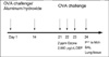
Female BALB/c mice 5-6 weeks of age were obtained from Daehan Laboratories (Daejeon, Korea). The mice were maintained on ovalbumin (OVA) free diets. The mice were individually housed in rack-mounted stainless steel cages with free access to food and water. An OVA-induced allergic airway disease model of asthma with some modification was used (17). Briefly, mice were sensitized by means of intra-peritoneal injection of on day 1, 14 day with 10 µg of Grade V OVA (Sigma Chemical, St Louis, MO, U.S.A.) and 1 mg of aluminum potassium sulfate (Sigma Chemical) in 500 µL of saline solution. Vehicle control mice were sensitized with a suspension of aluminum potassium sulfate (1 mg) in saline solution (500 µL) and challenged with nebulized saline solution daily from days 21 to 23 (Fig. 1). Aerosol challenge was carried out on groups of up 20 mice in a closed system chamber attached to an ultrasonic nebulizer (NE-UO7; Omron Corporation, Tokyo, Japan) with an output of 1 mL/min and 1- to 5-µm particle size.
DEP exposure
The engine used for preparation of DEPs was 4JB1 type (Isuzu Automatic Co., Tokyo, Japan), light-duty (2,740 mL), four-cylinder diesel engine. The engine was connected to an EDGY dynamometer (Meiden-Sya, Tokyo, Japan), was operated using a standard diesel fuel with speeds of 1,500 rpm under the load of 10 torque (kg/m). The exhaust was introduced into a stainless-steel dilutuion tunnel (300 mm Φ× 8,400 mm) in a constant-volume sampler system equipped to the end of the dilution tunnel. The temperature at the sampling point was below 50℃. The diameter of the particles was measured by an Anderson Air Sampler of the low-pressure type (18), and the mean diameter was 0.4 µm. Most of the shapes analyzed by a scanning electron microscope were globular. The details on the DEPs we used were previously described (19, 20). Mice were placed in exposure chamber and exposed to DEP 2,000 µg/µL for 1 hr and 2 ppm ozone for 3 hr after 1% (wt/vol) OVA challenge on days 21 to 23.
Ozone exposure
The mice housed in whole-body exposure chambers were exposed to ozone concentrations of 2 ppm for 3 hr (n=6/group) after 1% (wt/vol) OVA challenge on days 21 to 23. Ozone was generated with Sander Model 50 ozonizers (Sander, Eltze, Germany). The concentration of ozone within the chambers was monitored throughout the exposure with ambient-air ozone motors (Model 49C; Thermo Environmental Instruments Inc., Franklin, MA, U.S.A.). The air-sampling probes were placed in the breathing zone of the mice. The mean chamber ozone concentration (±SEM) during the 3 hr exposure period was 1.98±0.08 ppm. Temperature and humidity were maintained at a constant level within the chamber.
Determination of airway responsiveness to methacholine
Airway responsiveness was measured in unrestrained, conscious mice 1 day after the last challenge, as previously described (21). Mice were placed in a barometric plethysmographic chamber (All Medicus Co., Seoul, Korea), and baseline readings were taken and averaged for 3 min. Aerosolized methacholine in increasing concentrations (from 2.5 to 50 mg/mL) was nebulized through an inlet of the main chamber for 3 min. Readings were taken and averaged for 3 min after each nebulization and enhanced pause (Penh) was determined. Penh, calculated as (expiratory time/relaxation time-1)×(peak expiratory flow/peak inspiratory flow) according to the manufacturers' protocol, is a dimensionless value that represents a function of the proportion of maximal expiratory to maximal inspiratory box pressure signals and a function of the timing of expiration. Penh is used as a measure of airway responsiveness to methacholine. Results are expressed as the percentage increase of Penh following challenge with each concentration of methacholine, where the baseline Penh (after saline challenge) is expressed as 100%. Penh values averaged for 3 min after each nebulization were evaluated.
Bronchoalveolar lavage (BAL) fluid preparation and analysis
BAL was performed immediately after the last measurement of airway responsiveness. The mice were deeply anesthetized intraperitoneally with 50 mg/kg of pentobarbital sodium and were killed by exanguination from the abdominal aorta. The trachea was cannulated with a polyethylene tube through which the lungs were lavaged four times with 1.0 mL of physiologic saline (4.0 mL total). The BAL fluid was filtered through wet 4×4 gauze. Trypan blue exclusion for viability and total cell count was performed. The BAL fluid was centrifuged at 150×g for 10 min. The obtained pellet was immediately suspended in 4 mL of physiologic saline, and total cell numbers in the BAL fluid were counted in duplicate with hemocytometer (improved Neubauer counting chamber). Then, a 100 µL aliquot was centrifuged in a cytocentrifuge (Model 2 Cytospin; Shandon Scientific Co., Pittsburg, PA, U.S.A.). Differential cell counts were made from centrifuged preparations stained with Diff-quick, counting 500 or more cells in each animal at a magnification×1,000 (oil immersion).
RESULTS
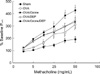
The OVA-sensitized-challenged and ozone and DEP exposure group had significantly higher methacholine-induced Penh than the sham group and the OVA-sensitized-challenged group (p<0.01, Fig. 2).
Numbers of total cells, the proportion of eosinophils, and neutrophils in BAL fluids were increased significantly in the OVA-sensitized-challenged and ozone and DEP exposure group than in the OVA-sensitized-challenged group and OVA-sensitized-challenged and ozone exposure group (Sham vs. OVA vs. OVA+Ozone vs. OVA+DEP vs. OVA+Ozone+DEP; eosinophils, 1.2±0.07% vs. 2.9±0.09% vs. 7.2±1.15% vs. 7.5±2.3% vs. 8.9±1.12%; p<0.05, neutrophils; 1.3±0.01% vs. 2.3±0.17% vs. 8.3±1.26% vs. 4.5±2.5% vs. 10.1±1.18%; p<0.05).
Level of IL-4 was increased in the OVA-sensitized-challenged and ozone exposure group and the OVA-sensitized-challenged and DEP exposure group, and the OVA-sensitized-challenged and ozone and DEP exposure group compared to the OVA-sensitized-challenged group (p<0.05, Fig. 3A). Levels of IFN-γ were significantly decreased in the OVA-sensitized-challenged and DEP exposure group and the OVA-sensitized-challenged and ozone and DEP exposure group compared to the OVA-sensitized-challenged and ozone exposure group (p<0.05, Fig. 3B).
DISCUSSION
The important finding of this study was that the methacholine-induced Penh, and IL-4, neutrophils and eosinophils in BAL fluids were increased in the allergen-sensitized-challenged and ozone and DEP exposure group than in the allergen-sensitized-challenged and ozone exposure group. These findings suggest that ozone and DEP may have additive effect on AHR in murine asthma model and the mechanism responsible for increased AHR by DEP and ozone exposure might be a contribution of Th2 immune responses.
Air pollution is a complex mixture of substances. Particles might exacerbate asthma that act indirectly by amplifying the allergic airway inflammation and increase directly airway responsiveness to common allergen. Ozone is a pollutant that coexists with particulates to a varying extent. We hypothesized that a co-exposure of DEP and ozone would exacerbate the airway inflammation and AHR in asthma patients. Therefore we evaluated the effect of co-exposure air pollutants on murine asthma model. Intranasal administration of DEP increased AHR to acetylcholine in both A/J and C57Bl/6 mice (22). The organic compounds of diesel exhaust particles (DEP-PAHs) caused AHR (23). The authors reported that Penh, an index of airway obstruction, was increased in a dose-dependent manner in mice exposed to ozone and persists for at least 72 hr (24, 25). In this study ozone have increased AHR to methacholine in the ozone-exposed group compared to that of OVA group. Also ozone plus DEP have more increased AHR indicating that DEP and ozone have additive effect of AHR.
Levels of IL-4 were increased significantly in the OVA-sensitized-challenged ozone exposure group and the OVA-sensitized-challenged DEP exposure group and the OVA-sensitized-challenged and ozone and DEP exposure group compared to the OVA-sensitized-challenged group. These results are consistent with those of Fujimaki et al. (26, 27), who reported that the adjuvant effect of DEP on IL-4 production and allergen induced IgE in mice challenged with allergen by the inhalation route, and that DEP effects on cytokine production in mice challenged intratracheally with 100 µg DEP for 6 wks: IL-2, IL-4, IL-5, and GM-CSF levels were increased compared with control animals. IFN-γ were significantly decreased in the OVA-sensitized-challenged and DEP exposure group and the OVA-sensitized-challenged and ozone and DEP exposure group compared to the OVA-sensitized-challenged and ozone exposure group. These results are consistent with the results with Diaz-Sanchez et al. that ragweed plus DEP challenge had a pronounced inhibitory effect on IFN-γ (28, 29).
Macrophages, eosinophils, neutrophils, and lymphocytes were significantly increased in BAL fluid from the animals challenged with DEP plus antigen compared to the animals treated with either agent alone (27, 30-32). DEP aggravated OVA-induced airway inflammation characterized by infiltration of eosinophils and lymphocytes and an increase in goblet cells in bronchial epithelium. In this study, eosinophils and neutrophils were increased in the OVA-sensitized-challenged and ozone and DEP exposure group compared to OVA-sensitized-challenged group, suggesting that eosinophils and neutrophils may play an important role in the pathogenesis of AHR of murine asthma model. Also DEP and ozone may be additive effect of airway inflammation.
The mechanism responsible for increased AHR of the DEP exposed to ozone is being speculative. Ambient concentration of ozone can increase the biological potency of DEP. The ozonized DEP may play a role in the induction of lung responses by ambient particulate matter (33). In this murine model of asthma co-exposure of ozone and DEP have increased AHR compared with that of ozone exposure, and increase of IL-4 and decrease of IFN-γ compared with that of ozone exposure indicating that balance of Th1/Th2 cytokine has a role in AHR of DEP exposed asthma model.
In conclusion, DEP and ozone have additive effect on AHR in a murine model of asthma via Th1/Th2 modulating activity.

 XML Download