Journal List > J Korean Med Sci > v.20(4) > 1020058

Bin, Kim, Kim, Keun, Lee, and Kim: Intrathoracic Aneurysm of the Right Subclavian Artery Presenting with Hoarseness: A Case Report

Abstract

Intrathoracic segment of the subclavian artery is an unusual location for peripheral arterial aneurysms. They are normally caused by atherosclerosis, medial degeneration, trauma, and infection. We report a case of a patient with right subclavian artery aneurysm presenting with hoarseness. Chest radiograph demonstrated a superior mediastinal mass. Laryngoscopy showed a fixed right vocal cord. By chest computed tomography, magnetic resonance imaging, and angiography, preoperative diagnosis was established as a saccular aneurysm with afferent loop and efferent loop. Patient underwent complete resection of the aneurysm followed by end-to-end anastomosis via median sternotomy. Postoperative pathology was consistent with an atherosclerotic aneurysm filled with thrombus. After surgical operation, hoarseness is still continued.

INTRODUCTION

Intrathoracic segment of the subclavian artery is a rare location for a peripheral arterial aneurysm (1-3). Intrathoracic segmental aneurysm is secondary to atherosclerosis, medial degeneration, trauma, and infection. Subclavian artery aneurysms are at an increased risk of rupture, embolization, or thrombosis. Hence, they should be considered for surgical repair. We describe a case of a patient presenting with hoarseness and an expanding intrathoracic mass that was due to a right subclavian aneurysm.

CASE REPORT

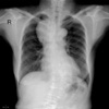
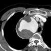
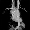
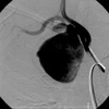
A 65-yr-old female presented with hoarseness that began 3 weeks ago. She had no previous history of trauma, pulmonary and bronchial tuberculosis. On admission, her vital signs were normal and blood pressure was equal on both arms. The laboratory workup showed a white blood cell count of 6,500/µL, normal erythrocyte sedimentation rate and C-reactive protein. Syphilis serology, rheumatoid factor, and antinuclear antibody tests were negative. Chest radiograph (CXR) demonstrated a superior mediastinal mass (Fig. 1). Because laryngoscopy showed a fixed right vocal cord, hoarseness was considered to be due to recurrent laryngeal nerve palsy. Computed tomography (CT) and magnetic resonance imaging (MRI) demonstrated a 7 cm sized proximal right subclavian artery aneurysm (Fig. 2, 3). Thoracic and subclavian aortography revealed a tortuous right subclavian arterial anatomy with a saccular aneurysm beginning near the subclavian origin (Fig. 4). The preoperative diagnosis was atherosclerotic right subclavian artery aneurysm.
Repair of the aneurysm was performed through a median sternotomy. We dissected at the aneurysmal afferent loop and efferent loop. We made end-to-end anastomosis by Gore Tex (6 mm sized) graft interposition. In the operation field, we could not find the recurrent laryngeal nerve due to severe adhesions. After the debridement, a partially undetached aneurysm was left.
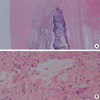
Cultures from the wall of the aneurysm and thrombus were negative. Pathology was consistent with an atherosclerotic aneurysm filled with thrombus (Fig. 5).
After one month follow-up, she still complains of hoarseness and laryngoscopic examination revealed right vocal cord palsy.

DISCUSSION

Aneurysms of the subclavian artery represent about 1% of all peripheral arterial aneurysms (4, 5). They fall into two distinct groups in terms of etiology, presentation, and treatment: those of the intrathoracic and those of the extrathoracic portion of the subclavian artery. Although aneurysms of extrathoracic subclavian artery are related to thoracic outlet syndrome or to previous trauma, intrathoracic segmental involvement is mainly due to atherosclerosis (6).
Intrathoracic aneurysms are most often asymptomatic but can present with symptoms caused by compression or acute aneurysm expansion such as upper chest or shoulder pain, Horner's syndrome, venous congestion, and hoarseness. Symptoms due to distal embolization to the arm are unusual. Extrathoracic aneurysm most commonly presents with a pulsatile mass in the superior fossa and is often tender, which may be noted by the patient or examiner. Brachial plexopathy is another complication of the extrathoracic aneurysm and Horner' syndrome is not infrequent (5-7).
Because isolated true aneurysm of the subclavian artery is rare, the natural history of subclavian artery aneurysm is unclear. It has been reported that an aneurysm of the aberrant right subclavian artery grew at the rate of 0.42 cm/yr measured by CXR (8). In another report, growth rate was reported at 1.31 cm/yr in a true left subclavian artery aneurysm measured by CT scan (9).
Elective surgical repair is the treatment of choice for most subclavian aneurysms, because they have an increased risk of rupture, embolization, thrombosis, and other complications. Intrathoracic subclavian artery aneurysms on the right are best approached by median sternotomy, whereas a high lateral thoracotomy is preferred for the left sided aneurysm (6, 10). Resection or aneurysmorrhaphy is preferred to simple ligation because continued growth and rupture of ligated aneurysm have been reported (11). Recently, as less invasive alternative to surgical repair, endovascular stent-graft treatment is possible (12-15).
We describe a case of a patient who complained of hoarseness and right shoulder pain due to an aneurysm of the intrathoracic subclavian artery. We treated her with graft interposition and left the aneurysm tissue unattached because of severe adhesion.

Figures and Tables

Fig. 1
PA chest roentgenogram shows well-defined, paratracheal mass of about 7 cm in the upper zone of the right hemithorax.
jkms-20-674-g001
Fig. 2
Contrast enhanced computed tomography (CT) demonstrates a 7 cm sized proximal right subclavian artery aneurysm and intraluminal yin-yang appearance due to thrombus.
jkms-20-674-g002
Fig. 3
Reconstructed three-dimensional magnetic resonance imaging (MRI) demonstrated positional relationships between right subclavian artery aneurysm and surrounding vessel.
jkms-20-674-g003
Fig. 4
Preoperative digital subtraction angiography of mass revealing an saccular aneurysm of right subclavian artery with afferent and efferent loop and intraluminal filling defects.
jkms-20-674-g004
Fig. 5
(A) Microscopically, atheromatous plaque contains amorphous pink material with slit-like "cholesterol clefts" of lipid material and calcification. There is recent hemorrhage on right side (H&E stain, ×20). (B) At higher magnification, many foam cells and a cholesterol cleft are seen (H&E stain, ×200).
jkms-20-674-g005

References

1. Chung YC, Jeong UG, Cho YK. A case report of subclavian arterial aneurysm. J Korean Surg Soc. 1978. 20:83–86.
2. Kim HJ, Kim SS, Huh JD, Chun BH, Joh YD, Cho SR. Intrathoracic aneurysm of the right subclavian artery. J Korean Radiol Soc. 1989. 25:725–727.
3. Kim HK, Kim KH, Park YS, Lee WH, Chug EC, Han WS. Subclavian artery aneurysm: Report of a case. Korean J Thorac Cardiovasc Surg. 1993. 26:557–559.
4. Dougherty MJ, Calligaro KD, Savarese RP, DeLaurentis DA. Atherosclerotic aneurysm of the intrathoracic subclavian artery: a case report and review of the literature. J Vasc Surg. 1995. 21:521–529.
crossref
5. Witz M, Yahel J, Lehmann JM. Subclavian artery aneurysms. A report of 2 cases and a review of the literature. J Cardiovasc Surg. 1998. 39:429–432.
6. Davidovic LB, Markovic DM, Pejkic SD, Kovacevic NS, Colic MM, Doric PM. Subclavian artery aneurysms. Asian J Surg. 2003. 26:7–11.
7. Utikal P, Bachleda P, Kocher M, Novotny J, Drac P, Drac P. Aneurysm of the subclavian artery. Acta Univ Palacki Olomuc Fac Med. 1999. 142:107–109.
8. Hogg JP, Dominic AJ, Counselman RL, Hurst JL. Expanding aneurysm of aberrant right subclavian artery. Case report and imaging evaluation. Clin Imaging. 1997. 21:195–199.
crossref
9. Takagi H, Mori Y, Umeda Y, Fukumoto Y, Yoshida K, Shimokawa K, Hirose H. Proximal left subclavian artery aneurysm presenting hemoptysis, hoarseness, and diplopia: repair through partial cardiopulmonary bypass and perfusion of the left common carotid artery. Ann Vasc Surg. 2003. 17:461–463.
crossref
10. Salo JA, Ala-Kulju K, Heikkinen L, Bondestam S, Ketonen P, Luosto R. Diagnosis and treatment of subclavian artery aneurysms. Eur J Vasc Surg. 1990. 4:271–274.
crossref
11. McCann RL. Basic data related to peripheral artery aneurysms. Ann Vasc Surg. 1990. 4:411–414.
crossref
12. Kim DK, Yoon YS, Choi SH, Lee DI, Lee DY, Chang BC, Shim WH. A case of transluminal stent-graft implantation at right subclavian artery pseudoaneurysm in Behcet's syndrome. Korean Circ J. 1999. 29:1240–1244.
crossref
13. Ko KH, Won JW, Won JY, Lee DY, No KS, Lee JT. Endoluminal placement of stent-graft for the treatment of peripheral saccular aneurysm. J Korean Radiol Soc. 2002. 46:213–219.
crossref
14. Schoder M, Cejna M, Holzenbein T, Bischof G, Lomoschitz F, Funovics M, Nobauer-Huhmann I, Sulzbacher I, Lammer J. Elective and emergent endovascular treatment of subclavian artery aneurysms and injuries. J Endovasc Ther. 2003. 10:58–65.
crossref
15. Kasirajan K, Matteson B, Marek JM, Langsfeld M. Covered stents for true subclavian aneurysms in patients with degenerative connective tissue disorders. J Endovasc Ther. 2003. 10:647–652.
crossref
TOOLS
Similar articles