

 PDF
PDF ePub
ePub Citation
 Print
Print


INTRODUCTION
Buckwheat is a common crops in Asia and Europe, and it is one of the most highly potent food allergens. Sensitization to buckwheat usually occurs by ingestion, but it can also occur by inhalation due to occupational or domestic exposure to flour (1-8). In 1909, Smith described a young patient who showed severe manifestations of asthma, allergic rhinitis, urticaria, and angioedema after ingesting of a small amount of buckwheat, the allergic symptoms were also induced by buckwheat inhalation (2). While there have been several related clinical reports on buckwheat allergy during the past 30 yr, a lack of epidemiologic studies has made it difficult to estimate the prevalence or incidence of allergic manifestations to buckwheat among populations in different countries.
In Korea, buckwheat flour is an important food allergen in school children and adults. Its positive skin test rate is about 5% (9). Several case reports have been issued on buckwheat hypersensitivity in Korea; two cases by the ingestion of cold noodle containing buckwheat flour (10), three cases by the inhalation of flour attached to a buckwheat husk pillow (11), and one case of occupational asthma by the inhalation of buckwheat flour in noodle making factory (12). Several allergenic components of buckwheat have been identified, and 24, 19, 16, and 9 kDa proteins are strong candidate major allergens in Korean allergic patients (13). Moreover, the 24 kDa β-chain of 11S globulin, has been identified a major allergen in Japan (14).
Buckwheat is one of the main dietary corps in Korea and Japan, furthermore, buckwheat consumption is increasing due to its nutritional benefits. Like peanut allergy, an allergic reaction to buckwheat is very severe and could be near fatal (2, 4, 15), but no satisfactory epidemiologic or mechanistic study has been reported, furthermore no effective therapy is available for this type of allergy. Consequently, animal models of buckwheat allergy, which mimic the immunological characteristics of buckwheat allergy in man, would be valuable tools for mechanistic research into grain allergies, such as, buckwheat, wheat and rice allergies. As far as we know, no study has been conducted on an animal model of buckwheat allergy. For this reason, we undertook this experiment with a view towards conducting further research.
There are several significant obstacles to the development of murine model of food allergy, such as, the strong innate tendency of animals to develop immunological tolerance to ingested antigens (Ag). Various studies in mice have shown that oral tolerance is influenced by strain (16, 17), age at first feeding (18-20), and the dose and nature of Ag (21, 22). In the present study, we employed a slightly modified approach of that used to generate a murine model of peanut-induced anaphylaxis by Li et al. (23). We demonstrated that immediate hypersensitivity reactions to buckwheat can be induced by intragastric sensitization and challenge. To validate the immunological relevance of this model to human buckwheat allergy, T and B cell responses to buckwheat extract, were evaluated by measuring serum antigen specific IgE, IgG antibody responses, splenocytes proliferation, and cytokine productions.
MATERIALS AND METHODS
Mice and reagents
Female C3H/HeJ mice, 5 weeks of age were purchased from the Jackson Laboratory (Bar Harbor, ME, U.S.A.) and maintained on buckwheat free chow, under specific pathogen-free conditions. Standard guidelines (24) for the care and use of animals were followed.
Fresh buckwheat flour was employed as an antigen for sensitization and challenge. Crude buckwheat extract was prepared as described previously (11). Cholera toxin (CT), concanavalin A (Con A), albumin was purchased from Sigma (St. Louis, MO, U.S.A.). Antibodies for ELISAs (Sigma) and ELISA kits to determine cytokine levels were purchased from the PharMingen (San Diego, CA, U.S.A.).
Intragastric sensitization and challenge
Mice were sensitized by intragastric (ig) feeding with fresh buckwheat flour solution on experimental days 0, 1, 2 and boosted on day 7. Intragastric feeding was performed by means of a stainless steel gavage needle, as described previously (23). To determine the optimum sensitization dose, mice were fedby ig with 1 mg (low dose), 5 mg (intermediate dose) or 25 mg per mouse (high dose) of buckwheat together with 10 µg/mouse of cholera toxin. Three weeks following this initial sensitization, mice were challenged ig with buckwheat at 10 mg/mouse divided in 2 doses at 30 min. intervals. The experiments were repeated twice using the same experimental protocol, and from the three experiments were controlled.
Assessment of hypersensitivity reactions
Anaphylactic symptoms were evaluated 30 min after the second challenge using the scoring system used in a peanut allergy model (23): 0-no symptoms; 1-scratching and rubbing around the nose and head; 2-puffiness around the eyes and mouth, diarrhea, pilar erecti, reduced activity, and/or decreased activity with an increased respiratory rate; 3-wheezing, labored respiration, cyanosis around the mouth and tail; 4-no activity after prodding, or tremor and convulsion; 5-death.
Measurement of buckwheat-specific IgE, IgG1 and IgG2a
Tail vein blood was obtained at weekly intervals following the initial sensitization. Sera were collected and stored at -80℃. Levels of buckwheat-specific IgE were measured by ELISA. Briefly, Immulon II 96-well plates (Dynatech Laboratories, Inc., Chantilly, VA, U.S.A.) were coated with 2 µg/mL crude buckwheate extract in coating buffer, pH 9.6 (Sigma). After an overnight incubation at 4℃, plates were washed and blocked with 1% BSA-PBS for 1 hr at 37℃. Following 3 washes, serum samples (1:10 diluted) were added to the plates and incubated overnight at 4℃. Plates were then washed 3× and 100 µL of biotinylated rat anti-mouse IgE (0.5 µg/mL) was added to each well. The plates were then incubated for 1 hr at 37℃, washed 3×, and 100 µL of avidin peroxidase (Sigma) (1:2,000 dilution) was added for an additional 30 min at room temperature. The plates were then washed 6× and ABTS (KPL, Gaithersburg, MD, U.S.A.) was added for 30 min at room temperature. The resulting reactions were read at 405 nm. Levels of IgE were calculated using a standard curve for measuring monoclonal antibody based on the ELISA method for total IgE (Sigma).
For determine PN-specific IgG1 and IgG2a levels, plates were coated with crude buckwheat extract and then blocked and washed in the same manner as described above. Samples (1:500 dilution) were added to the plates and incubated overnight at 4℃. The plates were then washed and biotinylated rat anti-mouse IgG1 or IgG2a monoclonal antibodies (1 µg/mL) were added to the plates to detect IgG1 and IgG2a, respectively. The plates were then incubated for an additional 1 hr at room temperature. After washings, avidin peroxidase was added for an additional 15 min at room temperature, and after an additional 8 washes, reactions were developed with ABTS for 15-30 min at room temperature and absorbance was read at 405 nm.
Proliferation assays
Spleens were removed from buckwheat sensitized and naive mice after re-challenge with buckwheat flour at week 5. Spleen cells were isolated and suspended in complete culture medium (RPMI 1640 plus 10% fetal bovine serum, 1% penicillin/streptomycin, and 1% glutamine). Spleen cells (1×10⁶/well in 0.2 mL) were incubated in triplicate cultures in microwell plates in the presence or absence of crude buckwheat extract (250 and 500 µg/mL). Cells stimulated with Con A (2 µg/mL) were used as a positive control. Three days later, the cultures were pulsed for 18-hr with 1 µ Ci per well of 3H-thymidine. The cells were then harvested and the incorporated radioactivity was counted in a β-scintillation counter. Results are expressed as ratios of counts per minute (cpm) of unstimulated- versus stimulated-wells.
Quantification of cytokines in splenocyte culture supernatant
Splenocytes were cultured in 24 well plates (4×10⁶/well/mL) in the presence or absence of PN (50 µg/mL) or Con A (2 µg/mL). IFN-γ, IL-4, and IL-5 levels in supernatants collected after 72 hr of culture were determined by ELISA according to the manufacturer's instructions (PharMingen).
Statistical analysis
Statistical analyses were performed using the Student's t-test for two group comparisons and using one-way ANOVA to compare more than two groups. A p value of <0.05 was considered statistically significant. All statistical analyses were performed using SigmaStat software (SPSS Inc. Chicago, IL, U.S.A.).
RESULTS
Systemic anaphylactic reactions following intragastric challenge
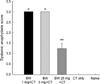
Three weeks following the initial sensitization, mice were fed crude buckwheat extract ig at 30-40 min intervals. Systemic anaphylactic symptoms were evident within 5-15 min of the first challenge. The severity of this anaphylaxis was evaluated 30-40 min after the first and second challenges. The initial reactions consisted primarily of cutaneous reactions, such as severe scrubbing around the whole body, puffiness, and cyanosis around the eyes and mouth, followed by labored respiration and decreased activity after prodding. Mice sensitized with the low dose (1 mg/mouse+CT) and medium dose (5 mg/mouse+CT) of whole crude buckwheat extract exhibited more severe reactions than those sensitized with the high dose (25 mg/mouse+CT). Fatal or near-fatal anaphylaxis did not occurred in this study. Sham-sensitized and naïve mice did not show any symptoms of anaphylaxis (Fig. 1). These results show that the initial sensitizing dose may influence the intensity of hypersensitivity reactions. We concluded that sensitization with buckwheat at the dose of 1 mg or 5 mg/mouse (low or medium dose) was optimal and that one of these doses should be used for subsequent study.
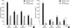
Buckwheat-specific antibody responses following sensitization and challenge
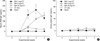
To explore the humoral immune responses underlying the development of buckwheat-induced hypersensitivity, sera from the different groups of mice were obtained weekly after ig sensitization and challenge. Levels of buckwheat-specific antibodies were determined by ELISA. Buckwheat-specific IgE concentrations were significantly increased from experimental week 1 through to week 4 in mice sensitized with low and medium dose groups, whereas the initial increment of buckwheat-specific IgE response was blunted after week 2 in the high dose group (Fig. 2A). Furthermore, buckwheat-specific IgE levels were consisted with systemic anaphylaxis symptom scores at week 3.
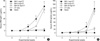
Buckwheat-specific IgG1 levels were also significantly different for the low/medium and high dose groups at weeks 3 and 4 (Fig. 3A). Similar to buckwheat specific IgG1 responses, IgG2a levels in the high dose group were significantly lower than in the low and medium dose groups from week 2 through to week 4 (Fig. 3B).
IgE responses to chow-protein extract
To determine IgE responses to daily-fed mouse chow protein extract, IgE antibodies against crude protein extract of mouse chow were measured by ELISA. Although, small chow-specific IgE responses were detected in the sham-sensitized group of mice after week 2, the levels of chow-specific IgE antibodies were negligible in all groups of mice (Fig. 2B).
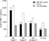
Splenocyte proliferative responses to crude buckwheat extract
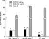
To characterize T-cell proliferative responses to crude buckwheat extract in this model, spleen cells from buckwheat allergic mice, sham sensitized mice, and naïve mice were cultured with crude buckwheat extract (50 and 250 µg/mL) at week 4 (Fig. 4). Although cells from all groups of mice, including CT control mice, showed significant proliferative responses to Con A stimulation, cells from buckwheat-sensitized mice, but not from sham-sensitized mice, exhibited significant proliferative responses to crude buckwheat extract. Unlike buckwheat specific IgE and IgG responses, antigen stimulated proliferative responses of splenocytes from the high dose group of mice were as strong as those from low dose mice.
Antigen specific T-cell cytokine responses
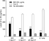
To determine the pattern of T-cell cytokine responses in this model, splenocyte cultures were performed with buckwheat extracts and Con A stimulations. As shown in Fig. 5, buckwheat specific IL-4 responses were significantly increased in the low-, medium-, and high-dose groups of mice, whereas antigen specific IL-5 responses were markedly increased only in the low-dose group. Furthermore, Con A seemed to have a slight stimulatory effects on IL-4 and IL-5 responses. Similar to Th2-type cytokine responses, buckwheat-specific IFN-γ responses were also predominant in buckwheat-sensitized mice, and were highest in the low-dose group. Moreover, the productions of IFN-γ were higher in the presence of Con A stimulation than in those with buckwheat stimulation (Fig. 6). Buckwheat specific IL-10 productions were also increased in buckwheat-sensitized mice, however in this model, IL-10 productions were markedly more induced by Con A stimulation than by antigen stimulation (Fig. 7).
DISCUSSION
Buckwheat sensitization occurs frequently by ingestion (1, 2, 10, 13, 14), and several related clinical reports have been published on buckwheat allergy during the past 30 yr, especially in Asia and Europe. However, a lack of epidemiological studies made it difficult to estimate prevalences or incidences. In Korea, buckwheat flour is one of the important food allergens in school children and adults, with a positive skin test rate of about 5% (9), and because of its nutritional advantages, buckwheat ingestion as a health food appears to be on the increase. Several case reports are available on buckwheat hypersensitivity in Korea (10-12), Japan (8), and more recently in Europe (15). As occurs in peanut allergy, allergic reactions to buckwheat are frequently severe and sometimes near fatal, furthermore some degree of cross allergenicity is suspected with wheat or rice, as was found during our previous studies (25). However, immunological and clinical studies on buckwheat allergy are lacking. Consequently, effective animal models of buckwheat allergy, which are capable of mimicking the immunological characteristics of buckwheat allergy in man, would be extremely valuable for mechanistic research into grain allergies, such as, buckwheat, wheat and rice allergies. As far as we know, this study is the first report about an animal model of buckwheat allergy. In the present study, we generated a murine model of buckwheat allergy induced by intragastric sensitization and challenge. Although in this study, the precise mechanisms involved in the sensitization at gastrointestinal sites, and systemic anaphylaxis following ingestion of buckwheat were unknown, observations of markedly increased buckwheat specific serum IgE and IgG1 antibodies, buckwheat stimulated splenocyte proliferations, and IL-4 and IL-5 production were definitely induced by the model.
It has been demonstrated that IgE plays an important role in mediating immediate type food allergy in humans (1, 26-28) and in animal models (23, 28, 29). In the present study, we found that buckwheat specific-IgE significantly increases through week 1 to 4-week following initial sensitization, especially in the low (1 mg/dose) and medium (5 mg/dose) dose sensitized groups of mice. Furthermore, buckwheat specific IgE levels at week 3 were consistent with symptom scores by intragastric buckwheat challenge at week 3. However, in this study, we could not confirm that the systemic anaphylactic symptoms stemmed from increased IgE levels, because we did not perform PCA reactions with positive sera. On the other hand, to determine IgE responses to daily-fed mouse chow protein extract, we measured IgE antibodies against crude protein extract of mouse chow by ELISA. We found that the levels of chow-specific IgE antibodies were negligible in all groups of mice through the experimental, which is a unique result that has not been discussed in other animal models of food allergy (23, 28). In the present study, to evaluate the differential effect of antigen doses during intragastric sensitization, we sensitized mice using three different doses of buckwheat extract. Interestingly, in the high dose (25 mg/dose) sensitized group of mice, the initial increment of buckwheat-specific IgE response was blunted after week 2, and buckwheat-specific IgE responses were significantly lower than this at week 3. Consistent with buckwheat-specific IgE levels, anaphylaxis symptom scores were lowest at week 3 among the three groups of buckwheat sensitized mice. Buckwheat-specific IgG1 responses showed patterns similar to those of IgE responses. Further studies are needed to evaluate the precise roles of both kinds of antibodies in this model.
To further assess the suitability of this model, we assessed T cell responses to buckwheat extract using a splenocyte culture system. We found significant proliferative responses to buckwheat extract stimulation in buckwheat-sensitized mice. Even though the degrees of splenocyte proliferations (presented by stimulation index) were highest in the cultures with Con A stimulation, relatively high (stimulation index: 7-10) levels of proliferations were observed in buckwheat stimulated cultures. Moreover, unlike buckwheat specific-IgE and -IgG responses, buckwheat stimulated proliferative responses in the high dose group of mice were as strong as those in the low dose group.
In addition, we confirm that buckwheat specific IL-4 responses were significantly increased in the low-, medium-, and high-dose groups of mice, and were highest in the low-dose group. However, antigen specific IL-5 responses were markedly increased only in the low-dose group. Furthermore, Con A seemed to have a relatively low stimulatory effects on Th2-type cytokines in this study. Similar to Th2-type cytokine responses, buckwheat-specific IFN-γ responses also predominated in buckwheat-sensitized mice, were highest in the low-dose group, and productions of IFN-γ were higher for Con A stimulation than for buckwheat stimulation. Buckwheat specific IL-10 productions were also increased in buckwheat-sensitized mice, but we could unearth either positive, or negative relations between IL-10 and levels of buckwheat specific IgE, or buckwheat stimulated IL-4 production. One important discussion point concerns the several possible effects of endotoxin contained in fresh crude buckwheat extract. Since studies have been performend on the fungal contamination derived endotoxin in storage grains, such as rice, wheat and buckwheat, and the direct effect of endotoxin on cytokine production by mouse T-cells (30, 31), we measured endotoxin levels in the crude buckwheat flour extract used in the present study. Its level was 616 EU/mg in our buckwheat extract, and thus we performed this experiment without eliminating endotoxin. Thus we could consider the possible effects of endotoxin on the production of all kinds of cytokines (i.e., IL-4, IL-5, IL-12, IL-10, IFN-γ) and on the proliferation of splenocytes in culture (31). However, fortunately, according to our experimental data, upon splenocyte proliferation and cytokine production, we found no additive influence of endotoxin on the proliferation capacities and cytokine productivities in naive or sham control mice. Thus we could use this model as a good quality of buckwheat allergy model in future study. And we need to repeat this experiment with endotoxin free or heated buckwheat extract in the future.
In conclusion, we established a mouse model of buckwheat allergy using intragastric sensitization and challenge, which closely reflects the clinical and immunological characteristics of immediate type food allergy in man. Buckwheat-induced allergic reactions in this model are IgE or IgG1-mediated, and buckwheat stimulated T-cell response up-regulation was observed. Using this model, we will extend our research on buckwheat allergy, for example, to identify and evaluate the major allergenic components of buckwheat extract, to confirm cross allergenicities with other crops, to test the capability of IgE induction without adjuvant during sensitization, and to develop therapeutic modalities for buckwheat allergy.
 XML Download