

 PDF
PDF ePub
ePub Citation
 Print
Print


INTRODUCTION
Whole body iodine-131 (I-131) scan remains the mainstay for the detection of recurrent or metastatic lesions of patients with well-differentiated thyroid cancer. The presence of uptake outside the areas of physiologic elimination such as salivary glands, nasopharynx, urinary bladder, and stomach is suggestive of metastasis. However, many false-positive findings on I-131 diagnostic and therapeutic scans have been reported (1-8). It is very important to recognize false positive findings to avoid unnecessary ablation treatment.
Previously, gallbladder localization of I-131 during post-therapy scan of well-differentiated thyroid cancer has been described (9, 10). However, no other study has reported normal gallbladder accumulation of I-131. We report a case of marked I-131 accumulation in normal gallbladder and this is the first report of normal gallbladder visualization in the absence of morphologic or functional abnormality (chronic cholecystitis, septated, or hypokinetic gallbladder) on post-I-131 therapy scan.
CASE REPORT
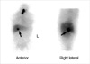
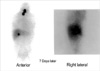
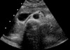
46-yr-old woman had a history of papillary thyroid cancer for which she had undergone total thyroidectomy. After total thyroidectomy, diagnostic whole body I-131 scan was performed. Forty-eight hours after the oral administration of a 111 MBq diagnostic dose of I-131 scan revealed thyroid remnants with regional lymph node metastasis and no abnormal focal hepatic accumulation. Blood analyses showed thyroglobulin <1.0 ng/mL and 10.2 U/mL of anti-thyroglobulin antibody. After a 4-week period of thyroid hormone withdrawal and limited ingestion of iodine containing foods, she was hospitalized for the radioiodine treatment for regional lymph node metastasis. She was received an ablative dose of 3.7 GBq of I-131. The whole body scans performed at 7 day showed areas of intense iodine accumulation in the thyroid bed and mild, diffuse hepatic uptake with a focus of markedly intense activity in the right hepatic lobe. In the right lateral of abdomen image, the iodine avid area in the liver was consistent with the location of gallbladder (Fig. 1). Another at 14 day whole body I-131 scan also showed sustained acitivity in gallbladder (Fig. 2). Abdominal ultrasound was performed and showed normal hepatic parenchyma and gallbladder (Fig. 3). There was no abnormal echo in gallbladder to suggest the presences of gallstone, inflammation, and metastasis.
DISCUSSION
Whole body I-131 scan is a well-established imaging method for the detection of metastatic or residual tumor sites in patients with well-differentiated thyroid cancer. However, there are many potential causes of false-positive I-131 scan findings (1-8). False positive results may be caused by a wide variety of non-thyroidal neoplasms, which can concentrate radioiodine or from skin contaminated by urine, sweat, or saliva. Several organs are capable of trapping iodine, including the salivary glands, gastric mucosa, choroids plexus, mammary glands, and renal cysts or pericardial effusion. Brucker-Davis et al. classified many false-positive findings into four main groups: elimination of iodine through body fluids, infection or inflammation, cyst or transduates, and non-thyroidal tumors (11).
Diffuse hepatic uptake of I-131 is a relatively common finding during whole body imaging. This uptake may reflect both I-131 liver blood pool activity as well as the physiologic hepatocellular concentration of I-131 labeled proteins and L-thyroxine (T4) produced by functioning thyroid tissue. The liver is major organ in the metabolism of thyroid hormone. It actively concentrates T4 and deiodinates it to T3. The liver is responsible for 40% of T4 deiodination occurring in the whole of the body and 70% of T3 production. Approximately 35% of exchangeable body T4 and approximately 5% of T3 are within the liver (12). The high incidence of diffuse hepatic uptake in cases of high doses of I-131 post-therapy scan can be explained by the fact that radiolabeled thyroid hormones are accumulated in the liver in patients with remnant thyroid tissues.
It is sometimes difficult to differentiate diffuse hepatic uptake and true hepatic metastasis. True hepatic metastases are typically seen as focal uptakes: the uptake by hepatic metastasis occurs earlier, with a time sequence similar to that of uptake by the thyroid or other metastases.
Thyroid hormones undergo metabolic degradation and excretion in conjugated form into the gut. Hence, I-131 uptake by the liver several days after I-131 administration is an index of the amount of functioning tissue present (8, 9, 13, 14). The gallbladder may occasionally be depicted when biliary excretion is extensive (8, 9).
Previously, gallbladder localization of I-131 during post-therapy scan has been described (9, 10). Two of these cases of patients had chronic cholecystitis. However, no other study has reported normal gallbladder accumulation of I-131 during post-ablative I-131 scan of thyroid cancer. According to Achong et al. (9), gallbladder visualization is likely a function of both the dose and the timing of the scan relative to I-131 administration. In our case, the gallbladder was seen 1 and 2 weeks after administration of high dose of I-131. However, it was not seen at 48 hr after low dose I-131 diagnostic scan. The gallbladder may have been seen if post-treatment I-131 scan had been performed at 48 hr instead of 1 and 2 weeks. Also, persistent gallbladder activity may be explained by abnormalities in both gallbladder morphology and functional dynamics such as septated hypokinetic and septated gallbladder with stone and/or chronic cholecystitis (15). Although, the functional study of gallbladder such as gallbladder ejection fraction measurement was not performed in this case, the ultrasound and clinical information did not show any morphological and underlying gallbladder disease.
This article documents the first report of I-131 radioiodine retention in the normal gallbladder that is not associated with gross structural and functional abnormalities. Although this is a rare finding, and the exact mechanism is unknown, it should be considered into the possible false positive findings of I-131 whole body scan.
 XML Download