

 PDF
PDF ePub
ePub Citation
 Print
Print


INTRODUCTION
Hamartoma is defined as an abnormal mixture of tissue elements, or an abnormal proportion of a single element, normally present in an organ. Pulmonary chondroid hamartoma (PCH) is the most common benign neoplasm in the lung, and accounts for 7-14% of all solitary lung nodules. However, cystic PCH is very rare (1-6). PCH of larger than 10 cm in size is also very rare (7).
We report a rare case of huge PCH with multilocular cysts in a 38-yr-old male patient.
CASE REPORT
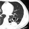
A 38-yr-old male presented with chronic cough and dyspnea for 8 months. Dyspnea was aggravated for the previous 4 days. Chest radiography showed a large multilobulated cystic and solid mass in the left lower lobe of the lung. Chest computed tomography revealed a huge multiseptated cystic and solid mass containing foci of intralesional calcifications in the left lower lobe of the lung (Fig. 1). A left lower lobe lobectomy was done under the impression of congenital cystic adenomatoid malformation (CCAM) or lung cancer.
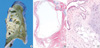
On gross examination, a huge cystic and solid mass containing variable size of multilocular cysts and solid component with numerous interstitial cartilaginous small nodules was found and occupied the superior segment and the upper portion of basal segment, measuring 11.5×10 cm in size (Fig. 2A). There was no connection with bronchus or vessel. Microscopically, multilocular cystic spaces with intervening lobulated fragments of cartilaginous tissue and hyalinized stroma were seen (Fig. 2B). The cysts and cleft-like spaces were lined by ciliated columnar epithelium. There were also foci of mature adipose tissues and a few spindle cells within the intervening stroma (Fig. 2C). Also seen were foci of calcification within the sclerotic stroma. The patient recovered uneventfully and there was no evidence of recurrence for nine months after the operation.
DISCUSSION
Hamartomas are the most common benign tumors of the lung and they comprise an admixture or overgrowth of various or single normal components that should be there. Depending upon the predominant component, hamartomas can be subdivided into various subtypes; chondromatous, leiomyomatous, lymphangiomyomatous, adenofibromatous and fibroleiomyomatous. Chondromatous hamartomas are the most common subtype and have been divided into endobronchial and intraparenchymal (peripheral) lesions. The onset of the tumor is in adulthood, with the peak age incidence in the sixth decade. Hamartomas may range from 1 to larger than 10 cm in the greatest dimension, but usually are smaller than 4 cm. One case has been reported of a tumor, measuring 16 ×9 cm in size (7). PCHs are frequently discovered on routine chest roentgenograms, in which they appear as solitary coin lesions. Less commonly, they may represent as multiple coin lesions or masses (8). However, even less frequently, cystic PCH may present as cavitary lesions on chest roentgenograms (5). In these hamartomas, cystic ones are very rare (1-6).
The mechanism of cyst development within a hamartoma is unknown. The route of entry of air into these lesions could be hypothesized, and check-valve mechanism might result in the gradual expansion of small epithelial-lined tubules resembling bronchioles (3). However, our case had no bronchial connection to the air-filled multicystic area. The growth condition of PCHs resulting from that the clefts-like spaces expanding to become growing cysts was also described (5).
Rearrangement of the high mobility group (HMG) proteins, non-histone DNA binding protein, HMGIC and HMCI (Y) has been recently proposed in PCHs (9). HMGIC-LPP (lipoma preferred partner) fusion gene has been described in two histologically different tumor types; lipomas and PCHs (10).
The differential diagnosis of cystic PCH includes CCAM, mesenchymal cystic hamartoma, cystic fibrohistiocystic tumors and cystadenoma. The CCAM is thought to be due to the arrest of the bronchial tree resulting in the adenomatous appearance composed of bronchiole-like or alveolar-like spaces. The mesenchymal cystic hamartoma is composed of cystic nodules with lining of normal or metaplastic respiratory epithelium and cambium layer of primitive mesenchymal cells without normal differentiation. The fibrohistiocystic tumors are presented as primary lung tumors or metastatic tumors of cellular dermatofibromas, which show frequent cystic change. The cystic lesions are associated with interstitial spindle cell proliferation, which demonstrates a storiform pattern, producing an overall impression of fibrous histiocytomas. The cystadenoma consisted of numerous irregularly arranged, tubular or dilated glands lined by a single layer of columnar, occasionally flattened, mucus-secreting cells. Hemorrhagic pattern of sclerosing hemangioma and cystic metastatic low-grade sarcomas are also included in differential diagnosis.
We may consider that the current PCH with huge size and multilocular cysts is exceptionally rare and should be differentiated from cystic pulmonary diseases.
 XML Download