

 PDF
PDF ePub
ePub Citation
 Print
Print


INTRODUCTION
Skeletal defects, derived from traumatic, degenerative, or tumorous causes, represent a major challenge to reconstructive surgery. For more complex bony defects, the conventional iliac crest non-vascularized corticocancellous free bone grafts have limited application and success. Despite their superiority as bone replacements and refinements in surgical technique, vascularized bone grafts or bone flaps, on the other hand, have not attained the popularity of soft tissue flaps. This is in part due to the limited donor tissue, donor site morbidity, complexity of the surgery, inability to shape the graft to the exact configuration of the bony defects and the availability of alternate. Alternative methods of skeletal reconstruction include the use of alloplastic materials or synthetic implants.
Since the early 1990s, significant progress has been made in constructing new tissue using cell transplantation. Transplantation of cells, instead of entire pieces of tissue, presents the opportunity to expand a small donor population into a larger transplantable mass. Furthermore, use of biodegradable polymer to deliver the cells and act as a templates for tissue regeneration may allow completely natural bone and cartilage (1, 2). Recently, there are many reports on bone engineering using periosteal derived cell as a cell source and various cell delivery vehicles such as a tricalcium phosphate (α-TCP), hydroxyapatitie (HA), polylactico-glycolic acid (PLGA) in Hutmachker's review (3). And also, prefabrication of vascularized bone graft using oseoconductive scaffold without cell transplantation have been reported (4).
We now report the construction of bone using periosteum derived cell-polymer constructs placed around existing vascular pedicles.
MATERIALS AND METHODS
Cell isolation, culture and characterization
Osteoblasts were isolated as described by Koshihara et al. (5). Periosteum was obtained from the humerus of newborn calf shoulders within 6 hr of sacrifice. Shoulders were rinsed in 10% povidone-iodine under sterile conditions, and the soft tissue was removed by sharp dissection. Periosteum was cut into pieces a 2×2 cm, rinsed in phosphate buffered saline (PBS), and placed into 35 mm tissue culture dishes (Costar: Cambridge, MA, U.S.A.). Each dish contains 2 mL of Media 199 (Gibco: Grand Island, NY, U.S.A.) supplemented with 10% fetal calf serum, L-glutamine (292 µg/mL), penicillin (100 U/mL), streptomycin (100 µg/mL), ascorbic acid (5 µg/mL) and Vitamin D3 (40 ng/mL). The specimens were incubated at 37℃ in the presence of 5% CO2 for 2 weeks to allow cells to migrate and proliferate in monolayer on the bottom of each well. One layer of cells was transferred onto a non-woven mesh of 15 µm polyglycolic acid fibers (Davis and Geck, Danbury, CT, U.S.A.), size of 5×7×1 mm. Cells adhered to and grew over the polymer mesh over the ensuing 7-10 days to form a multilayer of cells along the majority of the polymer fibers. Samples of the culture medium were analyzed for the presence of osteocalcin using a radioimmunoassay kit (Biomedical Technologies Inc. Stoughton, MA, U.S.A.). The results of this analysis were normalized for cell number by quantitating the number of cells adherent to the polymer scaffold. Culture media was exchanged once every 3 days.
Implantation of cell-polymer construct
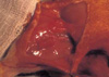
Cell-polymer devices were subsequently transplanted into athymic nude rats that weigh an average of 250-300 g under general anesthesia using inhalation of methoxyflurane and intramuscular injections of ketamine. Inguinal areas were washed with 10% povidone-iodine solution, and under sterile conditions, the femoral vessels of animals were exposed through a longitudinal skin incision under a surgical microscope. The polymer construct seeded with cells was wrapped around the right femoral vessel (experimental), and fixed in place with 6-0 nylon (Ethicon Inc. Piscataway, NJ, U.S.A.) suture (Fig. 1). The left femoral vessels were wrapped with polymer templates without cells (control). Three rats were sacrificed at 6 weeks, and nine rats were sacrificed at 9 weeks. Excised specimens were evaluated grossly and histologically by cutting 3 µm sections and staining with hematolxylin and eosin.
RESULTS
In vitro culture of cells
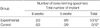
Cells readily adhered to the polymer fibers in multiple layers. To confirm that the isolated cells were osteoblasts, the secretion of osteocalcin, a protein specifically secreted by osteoblast, into the culture medium was quantitated. Osteocalcin was present in concentration similar to that previously reported (5) and concentration was upregulated by addition of vitamin D to the culture medium (Table 1).
Formation of new bone tissue in vivo
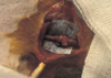
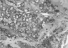
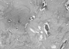
To assess the ability of these constructs to form vascularized bone tissue, they were subsequently implanted around the femoral vessels of athymic rats. Each polymer construct contained approximated 2×10⁶ cells at the time of implantation. Examination of excised specimens revealed progressive bone formation in the implants seeded with osteoblast (Fig. 2). The in vivo results are summarized in Table 2 and presented briefly below. Histological examination of the specimens revealed that progressive bone formation was found in 10 of 12 implants seeded with osteoblasts. Specimens at 6 weeks were primarily composed of cartilage with islands of osteoid tissue seen peripherally associated with blood vessel invasion (Fig. 3). At 9 weeks, the new bone was organized in that trabeculae were formed, and the bone appeared lamellar when sections were viewed using light microscopy (Fig. 4). However, control polymer devices implanted without cells did not result in bone formation over the entire time course. Grossly, there was no reminant of polyglycolic acid fiber in control side (left).
DISCUSSION
Free bone grafts are commonly used in reconstructive surgery. Use of vascularized bone grafts increase survival and minimize bone resorption. Experimental and clinical studies indicate that immediately vascularized autografts improved osteocyte survival and enhanced bone incorporation (6, 7). Thus, vascularized bone grafts or flaps are useful for patients with the poor recipient bed either scarred or irradiated.
To circumvent these limitations, our laboratory has been attempting to construct new structural tissues, such as bone and cartilage using cell transplantation (1, 8). By using synthetic biodegradable polymers as cell delivery vehicle it would be possible to engineer new, mature cartilage (9) and bone tissue in specific shapes and geometric design in near future. We have found that it is possible to engineer a vascularized bone by transplanting periosteum-derived cells around the pre-existing vascular pedicle using a biodegradable polymer as the cell delivery device. Degradation of the polymer over time leads to a nearly complete natural tissue, composed of autologous cells embedded within their own matrix and devoid of foreign materials. A central question arising from these results is whether the transplanted cell-polymer devices developed into bone tissue, or whether they induced host cells to migrate to the site and develop into bone tissue (10). The transplanted cells phenotypically resemble osteoblasts before transplantation, and critically, control polymers implanted without these cells induced no bone formation. These results suggest to us that the implanted cells were responsible for the new bone formation.
The results of these studies indicate that it is possible to create a vascularized bone tissue suitable for reconstruction using cells seeded on biodegradable polymer templates. Expansion of a limited source is possible with this technique, and this approach opens up the possibility of autologous cell transplants to reconstruct bony defects with vascularized bone grafts.

 XML Download