

 PDF
PDF ePub
ePub Citation
 Print
Print


Keratoacanthoma is a rapid growing hyperkeratotic papule occurring as a sporadic, solitary lesion on the sun-exposed skin of elderly individuals and usually undergoes spontaneous regression in a rapid growth over a 4-12 week period. Because it is difficult to differentiate between keratoacanthoma and squamous cell carcinoma in some cases, keratoacanthoma is generally considered to be a clinically and histologically distinct entity (1, 2). However, some reports have shown that keratoacanthoma is a type of squamous cell carcinoma with potential for aggressiveness and metastasis (3-5).
During tumor development, a normal cell is transformed to a malignant tumor cell via complex multi-stage processes. Neoplastic cells have undergone numerous genetic alternations, ranging from point mutations to chromosome aberrations, affecting the function or expression of both oncogenes and tumor suppressor genes. Loss of heterozygosity (LOH) is defined as a loss of genomic material in one of a pair of chromosomes. The LOH study is designed to assess polymorphic chromosome regions close to or within putative or known tumor suppressor genes. Thus, the LOH analysis can be used to obtain critical information on the discovery of new tumor suppressor genes such as Rb, BRAC2, NF1, and NF2 (6) and on the role of the presumptive tumor suppressor genes such as FHIT (7) in cancer development.
In the development of sporadic colorectal cancers, mutation of one allele of the APC tumor suppressor gene and consequent loss of the remaining allele are among the first genetic alternations (8). In the genetic progression of head and neck squamous cell carcinoma, studies on premalignant lesions have revealed LOH alternations as these premalignant lesions advance toward malignancy (9) and have indicated that LOH can take place in an early stage of tumorigenesis.
In this study, we analyzed 10 patients with sporadic keratoacanthoma to assess the potential role of LOH at six loci that previous studies displayed LOH in keratoacanthoma (3p25, 5q22-23, 9q32, 9p21, 10q23, and 17p12) (10-12) and at one locus known to have a specific breakpoint in a keratoacanthoma (2p12-13) (13) in the development of keratoacanthoma.
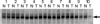
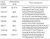
The study group was composed of 10 patients with sporadic keratoacanthoma (7 women, 3 men: 44-77 yr of ages, mean 63 yr). Cutaneous lesions were located on the nose (1 case), eyelid (4 cases), cheek (4 cases), and forehead (1 case). Samples were not collected if the lesion was recurrent, or if the patient was an organ transplantation recipient. Also patients with a history of prior chemotherapy or a family history of keratoacanthoma or visceral malignancies were not included. Unstained 5 µL thick tissue sections on glass slides were deparaffinized twice with xylene, rinsed with 95% ethanol, and stained with hematoxylin and eosin. Selected normal or tumor cell fields were microdissected under a light microscope using a 30 gauge needle and transfered to 100 µL of extraction buffer (10 mM Tris-HCl, 1% Tween, 0.1 mg/mL proteinase K, 1 mM EDTA, pH 8.0), respectively. The mixture was incubated overnight at 37℃ and boiled for 10 min to inactivate the proteinase K. The DNA solution was purified with QIA-quick Gel Extraction Kit protocol (Qiagen, Chatsworth, CA, U.S.A.). The purified DNA was eluted in 50 µL of TE buffer (10 mM Tris-HCl, 1 mM EDTA, pH 8.0) (14). LOH was examined by PCR using 7 microsatellite markers as listed in Table 1. PCR amplifications were performed with 4 µL purified DNA in a reaction mixture containing 0.3 µM of each primer, 0.2 mM of each dNTP, 15 mM MgCl2, 1 × reaction buffer, and 1.25 U AmpliTaq Gold™ DNA polymerase (Applied Biosystems, Foster City, CA, U.S.A.) in a final volume of 30 µL. PCR was performed for 35 cycles of 1 min at 94℃ for denaturing, 1 min at 55-60℃ for annealing of the primers, and 1 min at 72℃ for extension. Final extension was performed at 72℃ for 10 min. Five µL of each PCR product was added to 6 µL of formamide loading dye (95% formamide, 20 mM EDTA, 10 mM NaOH, 0.05% bromophenol, 0.05% xylene cyanol) and then denatured. Four µL of the denatured mixture was electrophoresed in 6.7% and 8% polyacryamide gel containing 7 M urea, respectively. Silver stain was performed to develop bands. Gel was fixed in 10% ethanol and 0.5% acetic acid for 6 min, then placed in 0.1% silver nitrate for 15 min and washed twice with distilled water. Reduction was processed in a solution containing 1.5% NaOH and 0.5% formamide until bands developed. The gel was placed in 0.75% Na2CO3 for 15 min, dried, and recorded. LOH was defined as positive when a clear reduction in signal intensity was detected in the tumors as compared with the corresponding normal alleles, or when a band corresponding to one allele of the normal DNA was lost in the tumor DNA.
The analysis of 10 sporadic keratoacanthomas matched with normal tissue samples revealed the presence of LOH at locus D10S185 (10q23) in only one out of the seven microsatellite loci examined and the frequency of LOH at that locus was found in only 1 of 10 keratoacanthomas. Fig. 1 demonstrates the electrophoretic pattern of the LOH positive keratoacanthoma (case 10) along with the pattern of corresponding normal tissue. No evidence of allele shifts meaning microsatellite instability was found at any of the tested markers in all cases.
LOH has been established as an important genetic mechanism giving rise to malignant neoplasia. Further more, the mechanisms of LOH have been observed to cause benign skin tumors. LOH loci at 9q34 and 16p13.3 in tuberous sclerosis harmatomas (15), at 9p22 in trichoepithelioma (16), at 16p13 in cylindroma (17), and at 1p and 9p in melanocytic nevus (18) have been found. Cutaneous tumors such as angiofibroma, collagenoma, or lipoma in patients with multiple endocrine neoplasia type 1 and neurofibromas in neurofibromatosis type 1 have been associated with allelic loss of MEN1 and NF1, respectively (19, 20). Therefore, the LOH studies have provided a view of genetic alternations to understand the pathogenesis of benign skin tumors.
The cytogenetic reports on keratoacanthoma are rare, but a karyotype with complex aberrations in a keratoacanthoma was reported in which chromosome rearrangements at both 2p13 were found (21) and we also presented a specific karyotype with 46,XY,t(2;8)(p13;p23) in a kearoacanthoma (13). Thus, the 2p13 region might be important in the pathogenesis of this skin tumor. In our study, although LOH at D2S286 (2p12-13) was not found, further other molecular studies at 2p13 in keratoacanthoma are needed to understand the biological characteristics of keratoacanthoma.
Muir-Torre syndrome is defined by the development in an individual of at least one sebaceous gland tumor and a minimum of one visceral malignancy, most frequently colorectal cancer. Additionally, further skin tumors including keratoacanthomas, basal cell carcinomas, squamous cell carcinomas, and actinic keratoses have been recorded in patients suffering from Muir-Torre syndrome (22). The APC tumor suppressor gene on chromosome 5 was analyzed because of its common involvement in gastrointestinal cancer (8). Our study and other two reports in keratoacanthoma (10, 11) could not detect LOH at D5S346, APC gene, suggesting that at this locus LOH may be not important in keratoacanthoma.
Waring et al. (12) showed LOH in 11 definite keratoacanthoma lesions. The examined loci were known frequently to be lost in squamous cell carcinoma of skin and in actinic keratoses (3p, 9p, 13q, 17p and 17q) and on other arms chosen at random. In that study, only two definite kearatoacanthomas showed LOH at three loci; one showed allelic loss on chromosome 9p (D9S162) and 10q (D10s185), and the other showed allelic loss on chromosome 9q (D9S160). In our study, LOH at chromosome 10q (D10S185) was found in 1 of 10 keratoacanthomas and therefore our results suggest that chromosome 10, at least the loci we examined, does not seem to play a major role in this tumor. And more cases of keratoacanthoma need to be analyzed to identify that LOH on chromosome 10q at D10S185 may be related to the pathogenesis of minor keratoacanthomas. On chromosome 3p, which region is thought to contain putative tumor suppressor genes, Waring et al. (12) did not found LOH at D3S1293 in 9 definite keratoacanthomas but detected LOH in 2 of 11 patients with probable keratoacanthomas. Peris et al. (10) observed LOH at D3S1317 in 2 of 20 patients with keratoacanthoma; one patient had a family history of visceral malignancies and the other had multiple keratoacanthomas. These findings, including our study, suggest that LOH at chromosome 3 is uncommon in sporadic keratoacanthomas and may appear to be importance in pathogenesis of advanced, aggressive keratoacnthomas or keratoacanthoma associated diseases such as Muir-Torre syndrome.
Chromosome 17 harbors a number of tumor suppressor genes including p53. Actinic keratoses are small, focal red area of cutaneous dysplasia with low risk of progression to squamous cell carcinoma. These lesions occur in sun-exposed skin, like as keratoacanthoma. High frequencies of LOH in actinic keratoses appeared on 17p (64%), 17q (56%), 13q (60%), 9p (31%), 9q (44%), and 3p (26%), contrasting with their benign clinical course (23). In keratoacanthoma, Langenbach et al. (11) observed not LOH but microsatellite instability at only D17S250 (p53) in 1 of 12 sporadic keratoacanthomas and Peris et al. (10) detected genetic alternations at p53 locus in 2 patients showing LOH at D3S1317 of 20 keratoacanthomas. One patient with multiple kerathoacanthoma showed LOH at 17S261 and the other patient with a family history of visceral malignancies, putative case of Muir-Torre syndrome, had multiple microsatellite instabilities including those at D17S261 and D17S520. We and Waring et al. (12) have detected no LOH at D17S261 and D17S796, respectively. Borkowski et al. (24) and Perez et al. (25) demonstrated frequency of p53 oncoprotein expression in nucleus by immunohistochemical staining was high, 77% (22/26) and 94% (15/16), respectively and suggested that p53 mutations contribute to the development of this benign tumor. These findings that no detection of LOH and presence of microsatellite instability at p53 locus in definite keratoacanthomas, and high frequency of p53 oncoprotein expression in keratoacanthomas suggest that the genetic alternations in p53 locus are not associated with LOH in the pathogenesis of this benign tumor.
Genetic alternations detected by LOH depend on the selection and number of chromosome markers that are chosen at chromosome regions. The relatively low level of LOH in keratoacanthoma thus may reflect the markers chosen. A comparative genomic hybridization study in keratoacanthoma detected high genetic aberrations showing deletions on 3p (20%), 9p (20%), 19p (20%), and 19q (16%) (26). Thus, more detailed LOH study on these chromosome regions is needed for evaluating LOH in keratoacanthoma.
In conclusion, our results suggest that LOH does not appear to be of importance in the pathogenesis of sporadic keratoacanthoma and other genetic events and genes are important in understanding the biological characteristics of keratoacanthoma.
 XML Download