

 PDF
PDF ePub
ePub Citation
 Print
Print


INTRODUCTION
Despite recent advancements in the diagnosis and treatment of systemic lupus erythematosus (SLE), infection remains the most important cause of death in this disease (1). Patients with SLE appear to be predisposed to an increased risk for infection due to immunosuppressive therapies as well as the intrinsic immunological defects associated with the disease (2). We report here on a patient with SLE who recovered from cryptococcemia and cryptococcal lymphadenitis two years ago, and the patient went on to develop mesenteric cryptococcal lymphadenitis even in the absence of any immunosuppressive treatment since the time of the original infection.
CASE REPORT
A 42-yr-old woman was admitted to Kangnam St. Mary's Hospital suffering from high fever and diffuse abdominal pain for three weeks. She had been diagnosed with SLE with class V lupus nephritis fourteen years ago. Two years ago, she had received corticosteroid and cyclophosphamide therapy for mesenteric vasculitis. After the treatment, she had fallen ill with cryptococcemia and retroperitoneal cryptococcal lymphadenitis. She recovered after her administration with treatment using intravenous fluconazole. However, she stated that she had not taken any medication for SLE such as corticosteroids and immunosuppressive agents.
On the physical examination, there was diffuse tenderness without rebound tenderness on her abdomen. There was neither arthralgia nor pitting edema on the extremities. A complete blood count revealed a mildly low hemoglobin level of 10.6 g/dL, a slightly elevated WBC count (10,800/µL with a differential count of neutrophils 70.4% and lymphocytes 20.2%), and a normal platelet count of 394,000/µL. The erythrocyte sedimentation rate (ESR) was 102 mm/hr and C-reactive protein (CRP) was 6.43 mg/dL. Her renal function was slightly impaired (creatinine 1.45 mg/mL, proteinuria 6.9 g/day and creatinine clearance 35.4 mL/min). The serum electrolytes showed normal levels. ANA was positive (1:160 with homogeneous pattern), anti-dsDNA antibody was 13.91 IU/mL (normal range 0-7 IU/mL), and C3 and C4 levels were in the normal range. Cryptococcal antigen was negative and there was no microorganism on the blood and stool cultures.
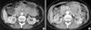
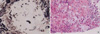
Abdominal computed tomography (CT) scan showed multiple conglomerated soft tissue density lesions noted in the mesenteric root, aortocaval space and left paraaortic space and smooth luminal narrowing was noted at the third portion of the duodenum: this resulted in the marked dilatation of the stomach and proximal duodenum (Fig. 1A). Under sonographic guidance, needle biopsy was performed at the conglomerated lymphadenopathies. Histopathologic finding showed chronic granulomatous inflammation with focal necrosis and the presence of fungal organisms, which were consistent with cryptococci (Fig. 2).
She was treated with intravenous amphotericin B 0.5 mg/kg/day, but after two weeks the serum creatinine was elevated up to 2 mg/dL, so amphotericin B was replaced with liposomal amphotericin B 1 mg/kg/day for eight weeks. This was followed by oral fluconazole 400 mg/day. Eight weeks after the liposomal amphotericin B administration, a follow up abdominal CT scan was performed. It showed marked decrease in the sizes and extents of the multiple conglomerated lymphadenopathies in the mesentery, and resolution of the previous noted obstruction of the duodenal third portion (Fig. 1B). The ESR and CRP were decreased to 38 mm/hr and 0.47 mg/dL. Oral fluconazole (200 mg/day) was maintained for twelve months and there was no sign of remained infection in clinical findings and last CT. For lupus nephritis, she was managed by angiotensin receptor blocker without any immunosuppressive agents due to the infection, and there was no change in renal function and proteinuria.
DISCUSSION
Cryptococcosis is an infection caused by a yeast-like fungus Cryptococcus neoformans. C. neoformans is an ubiquitous encapsulated yeast that can be isolated from soil and avian habitats, and most infections are thought to be acquired by inhalation of fungus into the lungs (3). Pulmonary infection has a tendency toward spontaneous resolution and this condition is usually asymptomatic. The organisms spread through the blood system and it mainly causes meningoencephalitis, but it can also affect other sites (4). Cryptococcosis due to C. neoformans is a common complication of late infection concurrent with HIV (5). Patients who have undergone solid-organ transplantation or glucocorticoid therapy are at increased risk for infections with C. neoformans (3).
This case is unique in that intraabdominal cryptococcal lymphadenitis occurred in the absence of immunosuppressive treatment, although the initial infection developed after immunosuppressive treatment. It is supposed that the intrinsic immune defects related to the SLE could have been responsible for cryptococcal lymphadenitis. Most cryptococcal infections present as meningoencephatlitis, followed by pulmonary and skin infection. Intraabdominal lymphatic cryptococcosis is very rare. There is only 2 reports of intraabdominal cryptococcal lymphadenitis in a patient with AIDS and omental cryptococcoma (6, 7).
The host defense mechanisms protecting the body against cryptococcal infections are complex and not completely understood. Cell-mediated immunity (CMI) is the crucial component of the immune system for resistance against cryptococcosis (8). This is supported by evidence that the most of the serious cryptococcal infections usually occur in individuals with defective CMI such as patients with acquired immunodeficiency syndrome (AIDS), corticosteroid treatment, reticuloendothelial malignancies or post-organ transplantation (9, 10).
Mok et al. have reported on a female SLE patient who developed cryptococcal meningitis concurrently with active SLE and in the absence of immunosuppressive treatment or other predisposing conditions (11). They suggested that her low complement level, low natural killer cell count and the probable defective CD4 positive T cell and macrophage functions resulting from her SLE disease activity might have been responsible for her susceptibility to cryptococcal meningitis.
A lot of immunological abnormalities that are present may either be the cause or the effect of SLE activity. These SLE related abnormalities include lymphopenia, defective CD4 positive T cell proliferation and interleukin 2 production to antigenic and mitogenic stimulation, reduced cytotoxicity of CD8 positive T cells, the impaired antigen-presenting function of monocytes and macrophages, hypocomplementemia, defective opsonization and the impaired neutrophil chemotaxis and phagocytosis (11, 12).
This case highlights that the importance of the intrinsic immunological defects of SLE predisposition to opportunistic fungal infection. To our knowledge, this is the first case report of mesenteric cryptococcal lymphadenitis occurred in a SLE patient without any immunosuppressive drugs.
 XML Download