

 PDF
PDF ePub
ePub Citation
 Print
Print


INTRODUCTION
Osteoporosis is defined as a skeletal disorder characterized by compromised bone strength predisposing a person to an increased risk of fracture (1). Bone mass accounts for approximately 70% of bone strength. At menopause, an uncoupling of bone remodeling, in which bone resorption exceeds bone formation, results in progressive bone loss and postmenopausal osteoporosis. It has been supposed that both the interleukin (IL) -1 family of cytokines and the IL-6 system may be involved in bone remodeling. IL-1α, IL-1β, and IL-6 are believed to be potent stimulators of bone resorption (2). Cytokine activity depends on the balance with natural modulators. IL-1 receptor antagonist (IL-1ra) is a competitive inhibitor of IL-1, and soluble IL-6 receptor (sIL-6r) acts as an agonist of IL-6. However, clinical studies on the possibility of involvement of these cytokines in the development of postmenopausal osteoporosis have led to contradictory results (3-9). Some of the conflict in results may be due to the differences in methodology of sample collections. A lot of advantages of whole blood cell culture in measuring cytokine secretion have recently been demonstrated, compared with serum measurement, or peripheral blood monocyte culture (10). Zheng et al. (11) reported that IL-1β and IL-6 production by whole blood cells was higher in osteoporotic postmenopausal women than in normal healthy women, but they did not adjust IL-1β production to total cell counts, and did not measure the level of IL-1α, IL-1ra, and sIL-6r. The balance between IL-1 and IL-1ra, and that between IL-6 and soluble IL-6 receptor are important to understand a whole picture of these IL systems' effects.
We have recently demonstrated that the cytosine-adenine (CA) polymorphism in the insulin-like growth factor-I (IGF-I) gene is associated with serum IGF-I levels (12). Similarly, polymorphisms within the cytokine genes may be genetic basis for the cytokine production by whole blood cells. Recently, a number of polymorphism in human IL-1 system and IL-6 gene have been reported: the C-889T polymorphism in the IL-1α gene (13), the C-511T polymorphism in the promoter region of IL-1β gene (14), the 86 base pair (bp) variable number tandem repeat (VNTR) in intron 2 of the IL-1ra gene (15) and the C-634G polymorphism in the promoter region of IL-6 gene (16) etc. The significance of these polymorphisms in terms of protein production by whole blood cells has not yet been determined. The aims of the present study were to investigate the relationship between the production of IL-1α, IL-1β??IL-1ra, IL-6, and soluble IL-6 receptor by whole blood cells, and bone mineral density (BMD) in postmenopausal Korean women and to evaluate whether IL system gene polymorphisms affect the production of ILs by whole blood cells.
MATERIALS AND METHODS
Study subjects
This study included 110 postmenopausal women, between the ages of 48 and 65 yr, who visited the Menopause Clinic of Seoul National University Hospital for bone mass examination, and who agreed to participate in the study. All of the subjects had been without spontaneous menses for at least one year. They underwent a careful physical examination, and a medical history review. Blood glucose, and hepatic and renal functions were determined. Women who had undergone bilateral oophorectomy or women with current hepatic disease, renal disease or diabetes mellitus were excluded. None of the subjects had received any medication known to affect bone metabolism before the study. All subjects gave their informed consent, and the study protocol was approved by the Institutional Review Board of Seoul National University Hospital.
Measurements of BMD and body mass index (BMI)
BMD was measured in grams per square centimeter at the lumbar spine (L2-L4), femoral neck, trochanter, and Ward's triangle, using a Lunar DPX-L dual-energy radiography absorptiometer (Lunar Radiation Corp., Madison, WI, U.S.A.), and categorized into three groups according to the World Health Organization's criteria (17) as, normal, osteopenic, or osteoporotic, relative to the means and standard deviation of young adult Korean women. The in vivo coefficient of variation was 1.4% for the lumbar spine, 2.1% for the femoral neck, 1.1% for the trochanter, and 2.1% for Ward's triangle. BMI was calculated by dividing body weight (kg) by the square of body height (m2).
Whole blood cells cultures
Blood samples were collected, from all subjects in accordance with the guidelines of the Declaration of Helsinki, with endotoxin free vacuum blood collection tubes containing preservative free heparin, and total number of monocytes and lymphocytes were counted. Blood was diluted 1/10 in RPMI (Gibco, Grand Island, NY, U.S.A.) complemented with 2 mM glutamine, 100 U/mL penicillin, 100 µg/mL streptomycin, 0.25 µg/mL amphotericin B and transferred into Falcon culture tubes with or without lipopolysaccharide (LPS) (Sigma, St Louis, MO, U.S.A.) at a final concentration of 25 µg/mL. Culture tubes were then incubated for 2 days at 37℃ in a 5% CO2 atmosphere. After incubation, samples were centrifuged at 800×g for 10 min, supernatants were collected, and frozen at -20℃ until assay for ILs.
Measurements of IL-1α, IL-1β, IL-1ra, IL-6, and sIL-6r in culture supernatant
IL-1α, and IL-1ra were measured using a quantitative sandwich immunoassay kit purchased from R & D systems (Minneapolis, MN, U.S.A.). The minimum detection limit was 1 pg/mL for IL-1α, and 14 pg/mL for IL-1ra. Intra- and interassay variations for IL-1α were 3.8% and 5.2%, respectively. Intra- and inter-assay variations for IL-1ra were 4.0% and 5.1%, respectively. IL-1β and IL-6 were measured using an enzyme linked immunosorbent assay kit purchased from Chemicon International Inc. (Temecula, CA, U.S.A.). The minimum detection limit was 0.195 ng/mL for IL-1β and IL-6. Intra- and inter-assay variations for IL-1β were 8.1% and 11.7%, respectively. Intra- and inter-assay variations for IL-6 were 7.0% and 11.3%, respectively. sIL-6r was measured using an enzyme linked immunosorbent assay kit purchased from Bender MedSystems (Vienna, Austria). The minimum detection limit was 0.02 ng/mL for sIL-6r. Intra- and interassay variations for sIL-6r were 1.7% and 2.2%, respectively. The resulting IL levels were then corrected for total monocyte and lymphocyte cell numbers. The values given are quoted in ng or pg/mL/106 cells.
Measurements of serum bone turnover markers
Serum osteocalcin (OST) was measured using a competitive radioimmunoassay kit (Techno Genetics, Milano, Italy), with a minimum detection limit of 0.1 nM/L. The intra- and inter-assay variations for OST were 4.0% and 5.1%, respectively. Serum carboxy-terminal C-telopeptide of type I collagen (CrossLaps; CTX) was measured using a serum CTX one step enzyme-linked immunosorbent assay kit (Osteometer Biotech, Herlev, Denmark), with a minimum detection limit of 94 pM/L, and intra- and inter-assay variations were 5.4% and 5.1%, respectively.
Determination of the IL-1 system and IL-6 gene polymorphisms
Genomic DNA was extracted from peripheral blood leukocytes using a QiaAmp blood kit (Qiagen GmbH, Hilden, Germany). The 86 bp VNTR polymorphism of the IL-1ra was analyzed as previously described (14, 15). A1 allele (four repeats) was 410 bp, A2 allele (two repeats) 240 bp, A3 allele (five repeats) 495 bp, and A4 allele (three repeats) 325 bp. The 304 bp fragment of genomic DNA containing the AvaI polymorphic portion at position -511 (C-511T) relative to the start site of transcription of the IL-1β gene was amplified by polymerase chain reaction (PCR), as described by Chen et al. (14). To analyze the NcoI polymorphic site at position -889 (C-889T) of the IL-1α gene, the 99 base fragment was amplified by PCR using the same oligonucleotide primers and PCR reaction steps previously described by McDowell et al. (13). To analyze the BsrBI polymorphic site at position -634 (C-634G) in the promoter of the IL-6 gene, PCR amplifications were carried out as described previously by Ota et al. (16). After PCR products were digested with restriction enzymes, the digests were separated on agarose gels. The absence of the AvaI, NcoI, and BsrBI sites was designated as the A, N, and B allele, respectively, and the presence of these restriction cutting sites was designated as a, n, and b allele, respectively.
Statistical analysis
All data are expressed as the mean±SE. Statistical analysis was performed using the SAS statistical program (SAS Institute, Cary, NC, U.S.A.). The genotype frequencies for each polymorphism against Hardy-Weinberg ratios were assessed by the χ2-test or Fisher's exact test. Differences in anthropometric characteristics between the different IL genotypes were tested using one way analysis of variance (ANOVA) followed by Tukey's test, or using Student's t-test. Similar comparisons were made for BMD, the serum levels of bone turnover markers, or for the production of ILs by whole blood cells using Student's t-test, or after adjusting potential confounding factors, such as age, and years since menopause, by analysis of covariance (ANCOVA). To assess the relationship between the production of ILs by whole blood cells and BMD, simple regression analysis was performed and Pearson's correlation coefficient was then calculated. A p value of less than 0.05 was considered significant for all analyses.
RESULTS
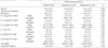
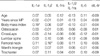
Table 1 shows the cytokine production by whole blood cells and serum levels of bone turnover markers in normal, osteopenic, and osteoporotic postmenopausal women. LPS treatment induced an increase in the production of IL-1α, IL-1β, IL-6, and sIL-6r by whole blood cells and an increased IL-1β/IL-1ra ratio. After adjusting for potential confounding factors, such as age, and years since menopause, no significant differences were observed in the basal production of IL-1α, IL-1β, IL-1ra, IL-6, or sIL-6r by whole blood cells, or in the IL-1β/IL-1ra ratio among normal, osteopenic, and osteoporotic postmenopausal women and this trend persisted even in the LPS stimulation. The production of IL-1 by LPS-stimulated whole blood cells correlated positively with BMD at the femoral neck (r=0.16, p<0.05), whereas the production of IL-1β, IL-1ra, IL-6, sIL-6r, and the IL-1β/IL-1ra ratio did not correlate with BMD at the skeletal sites examined (Table 2). There were no significant correlations between IL system components and bone turnover markers such as osteocalcin and CTX.
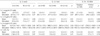
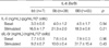
Four alleles in the IL-1ra VNTR polymorphism were observed: A1 96.4%; A2 1.8%; A3 1.4%; A4 0.4%. When study subjects were grouped according to carriage of the A2 allele, two genotypes were noted: the heterozygotes, A1A2, A3A2, (3.6%) and the noncarriage group, i.e., A1A1, A1A4, and A3A3 (96.4%). The distributions of the IL-1α NcoI, IL-1β AvaI, and IL-6 BsrBI polymorphisms were as follows; nn 85.5%, Nn 13.6%, NN 0.9%, aa 35.5%, Aa 50.9%, AA 13.6%, bb 6.4%, Bb 34.5%, and BB 59.1% respectively. These IL system allele frequencies followed the Hardy-Weinberg equilibrium. As shown in Table 3, regardless of LPS stimulation, the production of IL-1α, IL-1β, IL-1ra, IL-6, or sIL-6r by WBCs and the IL-1β/IL-1ra ratio were not different according to IL-1α, IL-1β or IL-1ra genotypes. IL-6 genotypes did not affect the production of IL-6, and sIL-6r by whole blood cells (Table 4).
DISCUSSION
A central role of immune system has been suggested in bone remodeling through proinflammatory cytokines. In the present study, we investigated the relationships among the production of interleukin-1 and IL-6 system by whole blood cells, and bone mass, and polymorphisms in IL-1 system and IL-6 genes in postmenopausal Korean women. To the best of our knowledge, this is the first report of its kind that have comprehensively analyzed about IL-1α, IL-1β, IL-1ra, IL-6, sIL-6r levels and IL-1α NcoI, IL-1β AvaI, IL-1ra A2, IL-6 BsrBI polymorphism.
It has been suggested that IL system may be implicated in the pathogenesis of postmenopausal osteoporosis. However, no differences has been noted in the circulating levels of IL-1α, IL-1β, IL-1ra (3), or IL-6 (4) or in the production of IL-1β by peripheral blood monocytes (5-6) between osteoporotic postmenopausal women and normal postmenopausal women, although some researchers have reported contradictory findings (7-9). Recently, Zheng et al. (11) reported that IL-1β and IL-6 production by mitogen-stimulated whole blood cells was higher in osteoporotic postmenopausal women than in normal postmenopausal women, and that lumbar BMD correlated negatively with IL-1β production. In the present study, regardless of LPS stimulation, we were unable to find any differences in the production of IL-1α, IL-1β, or IL-1ra, IL-6 or sIL-6r by WBCs, or in the IL-1β/IL-1ra ratios between osteoporotic postmenopausal women and normal postmenopausal women. A number of possible reasons may be proposed to explain the discrepancy of results between our study and that of Zheng et al. (11). First, they did not adjust the measured IL levels to total cell counts, whereas we did. Second, the whole blood cells culture time is relatively shorter in their study (1 day) than in our study (2 days), and it has been reported that the production of IL-1α and IL-2 reaches a maximum around 2-4 days after the start of whole blood cells culture (10). Third, they used both phytohemagglutinin and LPS, whereas we used LPS only. Finally, the number of study subjects in our study was twice as many as that from the report by Zheng et al. (11).
In the present study, IL-1β production by LPS-stimulated whole blood cells correlated positively with BMD of femoral neck. This finding suggests that high IL-1 does not always have a resorptive effect on all skeletal mass. This hypothesis is supported by recent findings that high cytokine production by peripheral blood monocytes is related with greater BMD gain in femoral neck, but greater bone loss in spinal bone (18). This possible site-specific skeletal effect of a certain cytokine needs to be elucidated by further studies.
Genotype frequencies of the IL-1 system genes in our subjects differ from those reported in Caucasians. The aa genotype of the IL-1β gene is less common than in Caucasians (35.5% vs. 43.4%) (19), and the NN the genotype of the IL-1α gene and A2 allele of the IL-1ra gene in Koreans were very rare, compared to Caucasians (13, 15). The distributions of IL-6 BsrBI genotypes in Koreans are similar to those previously reported in Japanese women (20). No differences were found in the production of IL-1α, IL-1β, or IL-1ra by whole blood cells, or the IL-1β/IL-1ra ratios among IL-1 system genotypes, although other investigators (18) have reported that the presence of the IL-1ra A2 allele is associated with an enhanced IL-1β and IL-1ra production, and a reduced IL-1α production by peripheral blood monocytes. These discrepancies may be due to differences in methodology of sample collection. In addition, there was no association of IL-6 genotypes with the production of IL-6 and sIL-6r in the present study.
Three types of sampling methods have been used to measure cytokine secretion; serum measurement, peripheral blood monocyte culture and whole blood cells culture. Of these methods, whole blood cells culture does not disturb the concentrations of circulating factors and the ratios of different cells, which may be important for normal molecular and intracellular interactions. Moreover, it has the advantage of being much quicker and simpler than peripheral blood monocyte culture, and is less likely to result in the contamination of samples.
In conclusion, IL-1β may regulate bone metabolism at femoral neck and IL polymorphisms do not affect the production of ILs by whole blood cells. Systemic ILs production by whole blood cells may not reflect the status of the cytokine production in the bone microenvironment. And thus, further cross-sectional and longitudinal studies are needed on the relationship between bone mass and cytokine production in the microenvironment of bone marrow.
 XML Download