

 PDF
PDF ePub
ePub Citation
 Print
Print


INTRODUCTION
Mature B-cell neoplasms, which are clonal proliferations of B cells at various stages of differentiation, comprise over 90% of lymphoid neoplasms in western countries and about 75% of non-Hodgkin's lymphomas (NHLs) in Korea (1-3). The distinctive morphology and immunophenotypes of mature B-cell neoplasms allow them to be readily classified by postulated cell of origin. Among many types of mature B-cell lymphoma, B-cell chronic lymphocytic leukemia (CLL) and mantle cell lymphoma (MCL) typically contain CD5 positive B cells, which can be observed within the period from naive B-cell to mantle cell. Recently, in addition to the two well-known lymphomas, another type of lymphoma with CD5 antigen expression has been reported in Japan (4, 5). This type lymphoma, so-called CD5 positive diffuse large B cell lymphoma (CD5+ DLBCL), was first described in western country, and has received much attention lately because it has been suggested to possess more aggressive clinical features and a poor outcome (6). Thus attempts have been made to categorize CD5+ DLBCL as new disease entity, distinct from CD5- DLBCL, CLL or MCL (7-9). The identification of a high-risk subgroup of DLBCL would be particularly important for determining therapeutic strategy, given that treatment is successful in only 35 to 40% of DLBCL patients (10). Therefore, we investigated DLBCL cases in all CD5 positive lymphoid malignancies with bone marrow involvement, as proven by flow cytometry, and also examined the clinical data, the histo-morphology, and immunophenotyping and cytogenetic result in these cases.
MATERIALS AND METHODS
We searched for CD5 positive cases of B-cell lymphoid malignancy involving the bone marrow among patients admitted to the Samsung Medical Center between 1997 and 2003. CD5 expression was confirmed by immunophenotyping using flow cytometry, which was performed using the following panel of lymphoid cell associated monoclonal antibodies: anti-CD2, CD3, CD5, CD7, CD10, CD19, CD20, CD23, FMC7, and surface immunoglobulin (Ig) (Becton Dickinson, CA, U.S.A.). The analyzed cells were gated on FSC/SSC and revealed lymphoid light scatter and moderate CD45 expression. Twenty-five cases of B-cell lymphoid malignancies showed CD5 expression; corresponding medical records were reviewed. Bone marrow histomorphologies of the 25 cases were re-evaluated using Giemsa and hematoxylin-eosin stains by three different pathologists. Diagnosis was based on the criteria documented in the REAL/WHO classification (11). In brief, CLL was defined as a case showing monomorphic small lymphocytes with CD5 and CD23 expression, and mantle cell lymphoma was restricted to cases with cyclin D1 expression by immunohistochemistry (Novocastra, CA, U.S.A.) in either a lymph node or bone marrow. Cytogenetic analysis was performed as follows: unstimulated bone marrow cells were cultured for 24 hr and G-banded, and twenty metaphase cells were analyzed and karyotyped, according to the guidelines of the International System for Cytogenetic Nomenclature (ISCN) 1995 (12). Dual color FISH with IGH/CCND1 probes was undertaken according to the manufacturer's instructions (Vysis Inc., Downers Grove, IL, U.S.A.), and 200 nuclei from interphase cells were analyzed. To determine the reference range of IGH/CCND1 FISH, 200 nuclei from 20 normal bone marrow specimens were observed; no true positive fusion signals (1G2O1F) were observed.
RESULTS
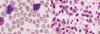
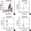
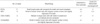
Among the 25 cases of B-cell lymphoid malignancies showing CD5 expression, 11 cases showed small sized lymphoid cells with clumped chromatin and scant cytoplasm expressing CD5 and CD23. These cases were diagnosed as B-CLL (Table 1). Another 9 cases showed small to medium sized cells with the characteristics of mantle cells; these were CD5+ and CD23-. Among these 9 cases, six expressed cyclin D1 by immunohistochemistry, but only two cases revealed the IGH/CCND1 rearrangement by FISH, and the remaining three cases showed neither cyclin D1 expression nor the IGH/CCND1 rearrangement. We finally diagnosed six cases showing cyclin D1 expression as MCL; the other three cases were unclassifiable. Finally, the remaining five cases showed medium to large-sized lymphoid cells with prominent nucleoli and a moderate amount of cytoplasm. Immunophenotypically they were positive for CD5, CD19, CD20 and surface Ig κ or λ, and negative for CD23 and CD10 (Fig. 1 and 2). All five cases were confirmed to be cyclin D1 negative, and a FISH study identified no case with the IGH/CCND rearrangement. After considering morphology, immunophenotype, immunohistochemistry, and FISH results, we ruled out CLL and MCL, and diagnosed the five cases as centroblastic type DLBCL with CD5+.
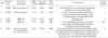
The characteristics of CD5+ DLBCL cases at presentation are summarized in Table 2. They showed a high age of onset (median, 68 yr), and no case had a history of low-grade lymphoma. Thus, they were categorized as de novo CD5+ DLBCL. They all received chemotherapy with the CHOP regimen (cyclophosphamide, doxorubicin, vincristine, and prednisone) and two patients (cases 1 and 2) expired one month after initial diagnosis.
DISCUSSION
The undefined three cases showing mantle cell morphology without cyclin D1expression and IGH/CCND1 rearrangement, which required further study including DNA microarray analysis, to define a diagnostic category. The presence of these cases implies that the REAL/WHO classification is not sufficient to classify diverse mature B-cell lymphoid malignancies. Recently, clusters of cases between typical CLL and MCL were reported based on flow cytometry, and they were found to be correlated with prognosis (13). Thus, an additional study should contribute to our understanding of mature B-cell lymphoid malignancies.
CD5+ cases account for approximately 5-10% of DLBCL, and usually the frequency of infiltration of bone marrow, the most frequent site of extranodal involvement, is reported to be around 25% (4, 5). In the present study, the frequency of CD5+ DLBCL among the 25 cases of CD5 positive B-cell lymphoid malignancies involving the bone marrow was 24%, which is not an insignificant number. In view of the fact that reports of CD5+ DLBCL are rare in Korea, a serious underestimation of CD5+ DLBCL is suspected.
De novo CD5+ DLBCL has been recognized to have distinct features; a high age at onset, a female preponderance, a frequent association with poor prognostic components, and an aggressive clinical course (5). Our cases also showed a higher average age of onset than general DLBCL in Korea (2) and frequent extranodal infiltration of both liver and spleen.
To explain the correlation between CD5 expression and an unfavorable prognosis, genetic aberrations of CD5+ DLBCL have been evaluated in a number of ways. Initially, an investigation targeting D13S25, p16, and p53 was performed, and the deletion at the D13S25 locus and loss of p16 were revealed, which implies an early transforming event resulting in aggressive DLBCL (8). In addition to the above mentioned genetic aberrations, the expressions of several other markers such as BCL2, cyclin D3, cyclin B, or survivin have been proposed to be associated with poor outcome (10). Following a DNA microarray analysis, it was reported that the outcome of patients with DLBCL might be influenced by gene expression profiles other than histologic profiles (14), and that the gene signature is associated with the control of cell kinetics and host immune response (15). Thus, study of the genetic aberrations of CD5+ DLBCL would help elucidate previously indistinct diagnostic categories.
The aggressive behavior of CD5+ DLBCL has also been attributed to a tendency to spread widely within tissue, and this phenomenon was associated with the CD5 molecule interacting as a ligand with heavy-chain variable framework regions of surface Ig (16). The latest study on this topic revealed that the aggressiveness of CD5+ DLBCL might be due to the overexpression of integrin β1 and/or CD36 adhesion molecules (17). Integrin β1, an adhesion molecule, plays an important role in B-cell lymphoma adhesion and chemotaxis on fibronectin (18). The protein expression of integrin β1 has already been reported to be associated with extranodal involvement in NHL (19). The CD36 antigen is transcriptionally regulated by Oct-2, which is a regulator of B-cell differentiation, and its expression is reported to be an indicator of tumor metastasis (20, 21). However, CD36 in CD5+ DLBCL tissues is expressed in the vascular endothelial cells rather than in malignant cells. This finding implies the pathophysiologic role of the vascular endothelium as an integral part of the cancer, and would explain the high frequency of intravascular or intrasinusoidal infiltration observed in de novo CD5+ DLBCL (5).
In conclusion, the present study found that de novo CD5+ DLBCL, with its distinct clinicopathologic features, is present in Korea. Moreover, the characteristics of our CD5+ DLBCL cases share many similarities with those previously reported. Since CD5+ DLBCL may form a distinct subgroup of DLBCLs, there is a need to determine the presence or absence of CD5 antigen expression in all DLBCL cases. It is thus essential to study CD5 expression in DLBCL by immunohistochemistry or flow cytometry, as such findings would be helpful for to predict prognosis and determine future therapeutic strategy. In addition, cytogenetic studies and molecular analyses for proto-oncogenes and tumor suppressor genes would provide insights into the pathogenesis of CD5+ DLBCL, and provide information relevant to the establishment of innovative therapeutic strategies. Furthermore, regarding the difficulty in making a diagnosis of heterogeneous mature B-cell lymphomas, comprehensive flow cytometry, cytogenetic and molecular genetic study would aid in the diagnosis of all mature B cell lymphoid malignancies.
 XML Download