

 PDF
PDF ePub
ePub Citation
 Print
Print


INTRODUCTION
Ophthalmoplegia is a rare but important complication of snake bites. It is developed by neurotoxins in snake venom. Neurotoxicity by snake venom is an indication of antivenom therapy. Although ophthalmoplegia is not a lethal condition following snake bites, the symptoms of diplopia and dizziness are emotionally devastating for patients and present diagnostic and therapeutic challenges to the physicians. Ophthalmoplegia by snake venom and its managements have rarely been reported in Korea (1, 2).
The authors describe two cases of ophthalmoplegia following snake bites, which did not improve with antivenom but rapidly improved by anticholinesterase therapy. These findings support the effectiveness of anticholinesterase in the management of ophthalmoplegia by snake venom.
CASE REPORT
Case 1
A 10-yr-old girl presented with a painful swelling of the right foot and diplopia. She was bitten on the toe about 6 hr prior to her admission by an unidentified snake inhabiting in Korea. She had felt pain and swelling of the bite site within several minutes, and was immediately taken to the hospital. By the time she arrived at the primary health care facility, local tissue edema was evident but systemic symptoms were absent. After a wound management without antivenom therapy, she was discharged. However, the pain aggravated and the swelling of the bite sites progressed above the ankle. After this time, the patient experinced new-onset diplopia.
On admission, she was alert and not so ill-looking. Initial vital signs were normal. Hemorrhagic blisters, bloody woozy, tender and erythematous swelling were noted at the bite site, while the edema was more expanded to the proximal joint. On eye examination, the visual acuity was 0.8/0.9 (OD/OS). The pupils were equal in size with normal reflex. She had right exotropia with a limitation of adduction (Fig. 1A). The right eye could not cross the midline on leftward gaze (Fig. 1A). The abnormal laboratory data included white blood cell count of 19,700/µL and serum creatine phosphokinase (CPK) 425 U/L (normal range, 51-246). After a careful interview and physical examination of the girl, we concluded that the diplopia was of new onset, and the snake venom induced the adduction deficit of her right eye.
An antivenom was infused immediately upon admission because the patient had newly developed diplopia and her pain and swelling aggravated. After the first infusion, her pain improved and the rate of swelling decreased, but diplopia persisted. The patient was treated with another dose of antivenom 4 hr after the initial dose, but still she complained of persistent diplopia. On the 2nd day of admission, she received intravenous (IV) neostigmine 0.5 mg per 6 hr and IV atropine 0.5 mg per 12 hr following a tensilon test. The signs of recovery from double vision and from a difficulty in focusing became evident after two injections of neostigmine. After 5 injections of neostigmine on the 3rd day of admission, the limitation of adduction in the right eye was completely recovered (Fig. 1B). She had a mild abdominal discomfort during the treatment of neostigmine, but it was self-resolved. The girl was discharged on the 8th day of admission without any ocular symptoms.
Case 2
A 13-yr-old boy was bitten by a snake on the right thumb about 18 hr prior to his admission. He visited our hospital because of a painful swelling of his right thumb and difficulty in focusing. He was immediately taken to the primary health care facility. After a wound management without antivenom therapy, he was discharged. However, the pain aggravated and the swelling of the bite sites progressed above the wrist. Headache and diplopia was newly developed at this time.
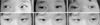
His initial vital signs were stable. On eye examination, the visual acuity was 0.9 (OU), and the pupils were equal in size with normal reflex. Diplopia aggravated with a limited motion of each eye on upward and inward gaze and on downward and inward gaze (Fig. 2A). Other eye examinations were normal. The laboratory data showed the prothrombin time was 13.2 sec (69.2%), activated partial thromboplastin time 32.1 sec, serum CPK 1,071 U/L, AST/ALT 78/95 IU/L, and urine myoglobin negative. We presumed that diplopia was of new onset because the snake venom had induced the limitation of ocular movement in each eye.
After the two doses of the antivenom, his pain improved, but the diplopia persisted. On the 2nd day of admission, he received IV neostigmine 0.5 mg per 6 hr and IV atropine 0.5 mg per 12 hr following a Tensilon test. The boy had a similar clinical course as in Case 1. After the 3rd injection of neostigmine, his ocular symptoms improved and after the 5th injection of neostigmine on the 3rd day of admission, the limitation of ocular motion was fully resolved (Fig. 2). We stopped neostigmine the day after the patient had full movement of EOM. The elevated levels of serum CPK and AST/ALT became normal without complications on the 3rd day of admission. He was discharged on the 5th day of admission without any ocular symptoms.
DISCUSSION
Venomous snakes inhabiting in Korea are Agkistrodon saxatilis, Agkistrodon blomhoffi brevicaudus and Agkistrodon calaginosus (1). The venoms of these snakes contain several enzymes and cyclolysin, hematotoxin, and neurotoxin (2). Although localized and systemic symptoms following snake bites are induced by cyclolysin and hematotoxin, many polypeptide neurotoxins cause muscle paralysis by blocking the nicotinic acetylcholine receptors at the post-synaptic motor endplates, or they affect the mode of neurotransmitter release at the presynaptic motor nerve endings (3).
Neurotoxic paralysis may also begin within the first hour of snake bites and is seen first as ptosis, then blurred vision and diplopia, followed by facial weakness and dysarthria. In severe cases, weakness of the limbs, paralysis of respiration, and fixed and dilated pupils may be observed (4). Our patients experienced blurred vision or diplopia. Ocular examination revealed adduction deficit in Case 1 and dysfunction of oblique muscles of each eye in Case 2. From the history of the present illness of the patient and also from the review of the literature, the snake bitten the girl and the boy would be Agkistrodon blomhoffi brevicaudus, known to be seen frequently in Korea. Among the medically important snake species of the Far East, bites by Agkistrodon blomhoffi usually do not cause serious effects (5). In some reports, the most common cause of eye symptoms following a snake bite is Agkistrodon blomhoffi (2). The most common eye symptom is ophthalmoplegia, and the medial rectus muscle is commonly involved. Also in our cases, the girl had paresis of the right medial rectus muscle; but the boy an had incomplete motion of each eye on upward and inward gaze and on downward and inward gaze, due to dysfunctions of oblique muscles, which is considered as a rare complication.
The main treatment of neurotoxic paralysis following snake bites is an injection of antivenom (6, 7). Indications for antivenom are hemostatic disturbance, cardiovascular abnormalities, neurotoxicity, elevated CPK and aminotransferases with a definite sign of local envenomation, and others (5, 7). We used a polyvalent anti-venom because it was difficult to identify species of the snake that had bitten the patients at the time of the treatment. Swelling, pain, and bloody woozing of the bite site were decreased following antivenom therapy, and high CPK and aminotransferases became normal over a short period. However, diplopia and ophthalmoplegia were not recovered.
To date, there is a lack of controlled studies evaluating therapy for ophthalmoplegia by snake venoms. Because the binding to the presynaptic portion is irreversible, clinical recovery occurs slowly and only with the formation of a new neuromuscular junction. The binding of toxin to the postsynaptic portion, however, may act postsynaptically to produce a competitive or noncompetitive acetylcholine receptor blockade (8). Although an antivenom may induce a certain degree of reversal of the paralysis by postsynaptic neurotoxin, the clinical recovery is, however, very slow. Anticholinesterase is potentially useful as in patients with suspected myasthenia gravis. Sung and Hah (2) reported a case of extraocular muscle paresis following a snake bite. Their case was the paresis of the left medial rectus muscle, and it took 14 days for the patient to recover from the paresis whereas it took only 3 days in our cases. They used only antivenom. The clinical response to anticholinesterase was more likely to ensue when the postsynaptic neurotoxin was predominant in the venom (9). All patients with neurotoxic symptoms should have a Tensilon test, and then the effects should be evaluated. If there is an improvement of symptoms or if there are no symptoms of cholinergic crisis such as muscle fasciculation, we recommend the use of anticholinesterase for patients with neurotoxicity following snake bites.
Although ophthalmoplegia is a rare symptom of snake bites, it may induce anxiety and ocular discomfort for a long period. Treatment of anticholinesterase with antivenom may facilitate the recovery from ophthalmoplegia.

 XML Download