

 PDF
PDF ePub
ePub Citation
 Print
Print


INTRODUCTION
Autosomal recessive limb-girdle muscular dystrophy (ARLGMD) is a clinically and genetically heterogeneous disorder affecting mainly the pelvic and shoulder girdle muscles. According to the genetic classification of ARLGMD, it has been divided into ten subtypes, from LGMD type 2A to LGMD type 2J (1). LGMD type2B (LGMD2B) is caused by mutations in the dysferline gene (DYSF, MIM*603009) located on chromosome 2p13.3, which induces a dysfunction of dysferlin at the protein level (2). Dysferlin is a member of the FER-1 protein family and contains six putative C2 domains, which can bind to phospholipids, inositol polyphosphates, Ca2+ and intracellular proteins (3). Dysferlin is expressed predominantly in the skeletal muscle and localizes to the plasma membrane of the muscle fibers. In addition, it has been suggested to be involved in membrane fusion (4, 5). A recent animal study has suggested that dysferlin plays a role in the sarcolemma repair process (6). The DYSF has been also shown to cause Miyoshi myopathy (MM), which is a rare form of distal myopathy with the calf muscles weakness. Identical mutations in patients with LGMD2B or Miyoshi myopathy have been noted in several reports, suggesting a role for the modifier gene(s) (7-11). Both LGMD2B and Miyoshi myopathy are referred to as "dysferlinopathy" because of the deficiency of the same protein.
Because of the clinical and genetic heterogeneity of the LGMD, the LGMD subtypes are classified based on genetic or protein analysis. The development of antibodies directed against dysferlin has recently been shown to be useful for making a diagnosis of LGMD2B by an examination of a muscle biopsy with immunostaining or immunoblotting techniques (4, 5, 10). In the current study, immunocytochemical examination for dysferlin was tested on a muscle biopsies from 17 Korean ARLGMD patients, and the clinical and pathological characteristics of four Korean LGMD2B patients, who showed the complete loss of the dysferlin protein on a muscle biopsy, were investigated.
MATERIALS AND METHODS
Patients Selection and Muscle biopsy
From March 2001 to November 2002, 17 unrelated, nonconsanguineous LGMD patients were enrolled in this study. The diagnosis of LGMD was established by the clinical history and physical examination, the family history, a normal nerve conduction study and the myopathic pattern on electromyography (12). All the patients underwent an open biopsy from the quadriceps femoris or biceps brachii muscle under local anesthesia. In all the cases, informed consents were obtained from the patients or their parents. Transverse serial frozen muscle sections (7 µm thickness) were stained with hematoxylin and eosin (H&E), modified Gomori trichrome, and a battery of histochemical techniques (NADH-TR, ATPase pH 9.4/4.6/4.3) were applied. All of them showed normal immunostaining for three domains of dystrophin (N-terminus, C-terminus and Rod-domain) on the muscle specimens, and the deletion was not found on multiplex PCR for selected exons of dystrophin gene.
Immunocytochemical staining of muscle specimens
The tissues were processed for immunocytochemistry as follows: the 7 m serial sections were fixed in acetone at 4℃ for 10 min, rinsed in 0.05 mol/L Tris-buffered saline (pH 7.5) for 15 min, and incubated for 30 min with a blocking solution containing 2% bovine serum albumin and 5% normal goat serum as described (13). The sections were then incubated overnight at 4℃ with one of the following antibodies: α-sarcoglycan (NCL-α-SARC, Novocastra), β-sarcoglycan (NCL-β-SARC, Novocastra), γ-sarcoglycan (NCL-γ-SARC, Novocastra), δ-sarcoglycan (NCL-δ-SARC, Novocastra), dysferlin (NCL-DYSF, Novocastra), β-dystroglycan (NCL-β-DG, Novocastra, Newcastle Upon Tyne, U.K.), and caveolin-3 (Transduction laboratory, Lexington, KY, U.S.A.). These affinity-purified antibodies were well characterized and did not cross-react with each other. After washing for 30 min in Tris-buffered saline, the sections were examined using secondary goat anti-mouse IgG antibodies conjugated to peroxidase and visualized by a DAB (diaminobenzidine)-peroxidase reaction (Vector Laboratories, CA., U.S.A.). The sections were also incubated with biotinylated goat anti-mouse IgG, which was followed by fluorescent isothiocyanate (FITC) avidin D (Vector Laboratory, CA., U.S.A.) for dysferlin.
RESULTS
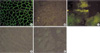
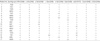
Four (Patient No. 1-4) of the 17 ARLGMD patients group (24%) showed a selective loss of dysferlin by both DAB and FITC methods on the muscle specimen (Table 1). Therefore, they were classified as having LGMD type 2B (LGMD2B). Although three patients (patient No. 8-10) showed isolated deficiency of δ-sarcoglycan, it was regarded as a technical error since the δ-sarcoglycanopathy had always showed combined deficiency of other sarcoglycans (α, β, and γ) at examination by immunocytochemistry (14). One patient (patient No. 15) showed loss of β and δ-sarcoglycan with partial deficiency of γ-sarcoglycan. This patient might be tentavely classified as β or δ-sarcoglycanopathy although molecular genetic diagnosis was not performed.
Clinical characteristics and course of disease in four patients with LGMD2B
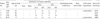
The age of patients at the onset of the disease varied from 9 yr to 33 yr, and the disease duration ranged from 6 yr to 15 yr (Table 2). All four patients showed symmetrical proximal weakness. Although all the patients showed a functional disability in walking or running, none was wheelchair-bound at the examination. While symmetrical hypertrophy of the calf muscle was noted in two patients (patient No. 1 & 4), muscle atrophy was not found in any of the four patients. None of the four patients had a cardiac or respiratory insufficiency nor did they have any bony abnormalities. The serum creatine kinase (CK) level was elevated to approximately 20 times of the normal level in all the patients. (4010-5310 IU/L). One patient (patient No. 1) had a positive family history. One of her elder brother (41 yr old) had similar symptoms, which presented as a slowly progressive proximal weakness over 15 yr.
Pathological and immunocytochemical findings of muscle biopsy in four patients with LGMD2B
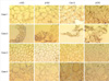
All the patients showed the following non-specific myopathic changes in the muscle: increased internal nuclei, a few or many atrophic fibers, degenerating fibers with regenerating fibers, and an increased fiber size variation (Fig. 1). Type 1 fiber predominance (>60%) was noted in one patient (patient No. 1). There was no endomysial or perivascular inflammatory cell infiltrations in all the patients.
DISCUSSION
The clinical features of our patients with LGMD2B was similar to those of previous reports (12, 15-17). Symptoms of the disease generally are manifested during the early adulthood (average onset is noted between the age of 17-30 yr), and the disease generally has a benign course, and confinement to a wheelchair may occur in approximately 10% of patients (15, 18). Despite of the slow clinical evolution, extremely high CK level (10-20 times of normal value) was characteristic of LGMD2B, being indicative of protein's role in normal muscle homeostasis ("leaky" membrane) rather than being essential for structural stabilization of the skeletal muscle (6, 19). A few LGMD2B patients showed symmetrical or asymmetrical calf muscle hypertrophy, as was observed in two of the patients in this study. Up to date, only several estimates of the frequency of dysferlinopathy exist. The proportion of LGMD2B is thought to be relatively high in Brazilian and Japanese population (estimated to be 19-25% of all ARLGMD cases) (15, 20) and there is founder mutation of DYSF appears to be present in the Libyan Jews (17). However, another recent study reported a relatively lower frequency of the dysferlin deficiency in Caucasian patients with the LGMD phenotype (approx. 1%), indicating a frequency variation according to the ethnicity (21). Although it is not enough to estimate the frequency of LGMD2B in the Korean ARLGMD patients due to a small number of subjects, these results suggest that the frequency of LGMD2B is relatively high among ARLGMD in Korea. A detailed population-based study will be needed to determine the exact frequency of the LGMD2B in Korea.
In all the cases with dysferlin deficiency, the immunocytochemical study showed normal reactivity against dystrophin, α-, β-, γ-, δ-sarcoglycans, β-dystroglycan on muscle specimen, which suggests that there are no interactions between dysferlin and the dystrophin-glycoprotein complex (DGC). This finding is in accordance with previous studies (22, 23). The DYSF is large, comprising 55 exons that span the genomic region of >150 kb (24). Although direct gene analysis provides the most reliable diagnosis, it is costly, time-consuming, and labor intensive because of the large size of the DYSF. Moreover, defects in the dysferlin gene involve mostly single nucleotide changes with no common mutations, gross rearrangements, or mutational hotspots that could aid detection (2). For this reason, it would be better to initiate screening for the dysferlin deficiency using antibodies against this protein in muscle biopsies. Complete loss of dysferlin without deficiency of other proteins appears to be specific for primary dysferlinopathy on immunocytochemistry (24). However, it should be noted that some cases with sarcoglycanopathy, caveolinopathy can show partial deficiency of dysferlin (secondary deficiency) (25, 26). Therefore, it is recommendable to perform immunocytochemical analysis using antibodies against other sarcolemmal proteins in addition to the antibodies against dysferlin. When partial deficiency of dysferlin is present on immunocytochemistry, direct sequencing of DYSF will be needed.
In conclusion, the clinical, pathological, and immunocytochemical findings of patients with LGMD2B in this study were in accordance with those of previous reports.

 XML Download