

 PDF
PDF ePub
ePub Citation
 Print
Print


INTRODUCTION
Perinatal hypoxic-ischemic brain injury remains a major cause of neonatal and infant mortality, and of permanent neurodevelopmental sequelae such as mental retardation, seizure disorders and cerebral palsy (1). Hypoxic-ischemic brain damage is an evolving process, which begins during the primary hypoxic-ischemic insult and extends into the recovery period after oxygenation and perfusion have been restored (2). Evidence is accumulating in neonatal brain ischemia models that the post-ischemic reperfusion period may be of major pathogenetic importance (3). It has subsequently been confirmed that ischemia-reperfusion in other species, including rat, leads to production of free radicals (4). These reactive oxygen species and their product, lipid peroxides, are thought to be among the important causes of cell membrane destruction and cell damage (5, 6).
There have been attempts to eliminate free radicals through inhibition of xanthine oxidase with allopurinol (7, 8). However, the results were mostly negative, probably because allopurinol does not penetrate well into the brain and because inhibition of xanthine oxidease there is minor and incomplete (9). Free radical scavengers such as superoxide dismutase and catalase have been shown to ameliorate the ischemic brain damage (10, 11). However, the therapeutic potentials of these free radical scavengers are limited by circulatory half-lives of only 6 to 10 min following intravenous injection (12). Furthermore, partially reduced oxygen species can only diffuse short distance before reacting with cellular components, and neither superoxide dismutase nor catalase can penetrate cell membrane to gain access to intracellular sites of free radical generation (13).
The spin trapping agent, α-phenyl-n-tert-butyl nitrone (PBN) is a well-recognized tool with which to demonstrate free radicals. It reacts with reactive oxygen species to form stable adducts that can be detected, identified, or quantitated (14, 15). Recently, PBN has been discovered to have a neuroprotective effect on focal ischemic brain injury. PBN reduced the infarct size and prevented the secondary energy failure and mitochondrial dysfunction (16-18). Although the detailed mechanism of its action is unclear, the neuroprotective effect of PBN seems to result primarily from efficient scavenging of oxygen free radicals (19). In addition, PBN has a low toxicity, and because of its lipophilicity, this substance readily crosses the blood-brain barrier, and penetrates well into the cell membranes (20, 21). These features make PBN an attractive therapeutic agent to ameliorate the brain damage from hypoxic-ischemic brain injury.
However, there are few reports about the effect of PBN on global hypoxic-ischemic injury in developing brain (22). This study was done to determine whether free radicals mediate brain injury during hypoxia-ischemia and reoxygenation-reperfusion, and whether the brain injury is attenuated by PBN in newborn piglets. We tested the hypothesis that PBN attenuates brain damage by scavenging free radicals during hypoxia-ischemia and reoxygenation-reperfusion of perinatal asphyxia. In this study, we used the newborn piglets as an animal model of perinatal hypoxic-ischemic brain injury because the piglet brain is comparable in growth velocity to human brain at birth. Changes in brain cell membrane structure, function, and energy stores during hypoxia-ischemia and reoxygenation-reperfusion were determined by measuring lipid peroxidation products (conjugated dienes), Na+, K+-ATPase activity, and concentration of high-energy phosphate compounds in the cerebral cortex, respectively.
MATERIALS AND METHODS
Animal preparation and surgical procedures
The experimental protocols described herein were reviewed and approved by the Institutional Animal Care and Use Committee of the Samsung Biomedical Research Center, Seoul, Korea. This study also followed the institutional and National Institutes of Health guidelines for laboratory animal care.
Newborn piglets less than 3 days old and of mixed strain (Yorkshire, conventional breed, purchased from Paju farm, Paju, Kyonggi-Do, Korea) were used in this study. Animals inhaled ether for sedation, and anesthesia was induced with thiopental sodium (5 mg/kg, i.v.), and supplemental doses were given when necessary to maintain anesthesia. After local injection with lidocaine (1%), a tracheostomy was performed and the piglet was paralyzed with pancuronium (0.1 mg/kg, i.v.) and ventilated with neonatal pressure-limited, time-cycled mechanical ventilator (Sechrist Infant Ventilator, IV-100 V, Sechrist Industries, Anaheim, CA, U.S.A.). Ventilator settings were adjusted to keep the arterial partial oxygen pressure at 80-150 mmHg and the arterial partial carbon dioxide pressure at 35-45 mmHg. Femoral artery and vein were cannulated for monitoring blood pressure, blood sampling, and for medication and fluid infusion. Bilateral common carotid arteries were isolated at the level of the fourth cervical vertebral level and encircled with silk surgical thread (size 4). Throughout the experiment, the piglet was placed under the servocontrolled warmer (Airshields, Hatboro, PA, U.S.A.), and rectal temperature was maintained between 38.0 and 39.0℃.
Experimental protocol
Fifty-one newborn piglets were divided randomly into the following four experimental groups: nine in the sham operation control group (C); 13 in the hypoxia-ischemia with vehicle group (HI-C); 10 in the hypoxia-ischemia with PBN group (HI-PBN); 9 in the reoxygenation-reperfusion with vehicle group (RR-C); and 10 in the reoxygenation-reperfusion with PBN group (RR-PBN).
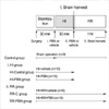
The experimental protocols for each group are described in Fig. 1. The total duration of each experimental treatment was 1.5 hr after obtaining stabilization. After surgery and stabilization, all animals were ventilated to maintain normal blood gas levels. In C group, normoxic ventilation was maintained for 1.5 hr. In hypoxia-ischemia groups (HI-C, HI-PBN), acute global cerebral hypoxia-ischemia was induced by temporary but complete occlusion of bilateral common carotid arteries with surgical clips and simultaneous breathing with 8% oxygen for 30 min. In reoxygenation-reperfusion groups (RR-C, RR-PBN), acute global hypoxia-ischemia was induced with the same method as in hypoxia-ischemia groups, and after then, the carotid artery occluders were released, inspired oxygen concentration was increased to maintain oxygen saturation at 90-95%, and normoxic ventilation was continued for one hour.
In PBN treatment groups (HI-PBN, RR-PBN), 100 mg/kg of PBN (Sigma Chemical CO., St. Louis, MO, U.S.A.) in normal saline (10 mg/mL) was given as a bolus intravenous injection just before the induction of hypoxia-ischemia (HI-PBN) and reoxygenation-reperfusion (RR-PBN), respectively. In PBN control groups (HI-C, RR-C), vehicle (saline) was given in the same way as in PBN treatment groups. Heart rate, oxygen saturation and blood pressure were monitored continuously during the experiment using the Hewlett-Packard neonatal monitoring system (Hewlett-Packard Model M1276A, MA, U.S.A.). Arterial blood gases and concentrations of glucose and lactate in the blood were measured at baseline, the end of hypoxia-ischemia, and every 30 min after hypoxia-ischemia.
At the end of each experiment, whole brain tissue was harvested immediately using guillotine, frozen rapidly in liquid nitrogen, and stored at -80℃ for further biochemical analysis. Arterial blood gases were measured on a blood gas analyzer (Ciba-Corning) and concentrations of glucose and lactate were measured using a YSI model 2300 dual analyzer (Yellow Springs Instrument Co., Yellow Springs, OH, U.S.A).
Biochemical analyses of brain cortex
Methods of brain cell membrane preparation and determination of cerebral cortical cell membrane Na+, K+-ATPase activity, levels of conjugated dienes, tissue glucose and lactate concentrations, ATP and phosphocreatine were described in detail previously (30). Briefly, brain cell membranes were prepared according to the method described by Harik et al. The activity of Na+, K+-ATPase was determined by subtracting the enzyme activity in the presence of ouabain from the total activity in the absence of ouabain. The level of conjugated dienes was determined using the method of Recknagel and Glende. The concentrations of glucose and lactate in the cerebral cortex were determined spectrophotometrically using a commercial kit (Sigma Chemical Co., St. Louis, MO, U.S.A.). Brain ATP and phosphocreatine levels were determined with a coupled enzyme assay using the method of Lamprecht et al.
Statistical analysis
Data were analyzed by one-way ANOVA or Kruskal-Wallis test for intergroup comparisons. To detect significant changes over time within each group and between groups, data were compared using repeated measures ANOVA with Bonferroni post-hoc-test. Statistical analyses described above were done using SPSS version 11.5. A p-value of <0.05 was considered significant. Data were given as mean±standard error.
RESULTS
Physiologic variables
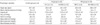
After hypoxia-ischemia was induced, heart rate significantly increased, and mean arterial blood pressure significantly decreased. During reoxygenation-reperfusion, mean arterial blood pressure returned to near its pre-hypoxia-ischemia level, while heart rate remained at as high levels as during hypoxia- ischemia. Arterial pH, base excess, and PaO2 significantly decreased during hypoxia-ischemia. During reoxygenation-reperfusion, PaO2 recovered to its pre-hypoxia-ischemia level, while arterial pH and base excess remained at as low levels as during hypoxia-ischemia. PaCO2 did not change significantly throughout the experiment. The concentrations of glucose and lactate in the blood both significantly increased after hypoxia-ischemia was induced, and then they did not change significantly to the end of one hour of reoxygenation-reperfusion (Table 1).
PBN did not influence above physiologic variables during hypoxia-ischemia and during reoxygenation-reperfusion (Table 1).
Biochemical data in brain cortex
After hypoxia-ischemia was induced, brain lactate level significantly increased, and recovered halfway during reoxygenation-reperfusion, while brain glucose level did not change significantly all through the experiment. The brain cortical tissue level of lipid peroxidation products (conjugated dienes), measured as an indicator of alterations in cell membrane structure significantly increased during hypoxia-ischemia, and remained elevated during reoxygenation-reperfusion. Na+, K+-ATPase activity in the cerebral cortical cell membrane, measured as an index of brain cell membrane function and cerebral ATP and phosphocreatine concentrations, measured as an index of cellular energy state were both significantly decreased after hypoxia-ischemia was induced. During reoxygenation-reperfusion, cerebral ATP and phosphocreatine concentrations recovered to near their pre-hypoxia-ischemia levels, while Na+, K+-ATPase activity remained decreased as during hypoxia-ischemia (Table 2).
PBN significantly decreased the level of conjugated dienes both during hypoxia-ischemia (0.99±0.09 vs.1.09±0.10) and during reoxygenation (0.96±0.10 vs.1.06±0.07). PBN also decreased brain lactate level markedly during reoxygenation-reperfusion, however the decrease was not statistically significant. PBN did not make any significant changes in brain glucose level, Na+, K+-ATPase activity, and ATP and phosphocreatine concentrations during hypoxia-ischemia and during reoxygenation-reperfusion (Table 2).
DISCUSSION
In the present study, the level of lipid peroxidation products (conjugated dienes) in the brain significantly decreased when newborn piglets were pretreated with PBN not only during reoxygenation-reperfusion, but also during hypoxiaischemia. Because lipid peroxides are products of reactive oxygen species, this result shows that PBN acted as an effective free radical scavenger in our piglet model of hypoxiaischemia and reoxygenation-reperfusion. Membrane lipid peroxidation is among the important causes of cell membrane destruction and cell damage, and is an indicator of the amount of oxidative stress related to secondary energy failure (5, 6, 24, 25). Secondary energy failure has been known to be associated with poor neurodevelopmental outcome (26). Therefore, it is suggested that PBN may be neuroprotective in hypoxic-ischemic brain injury.
During hypoxia-ischemia, there is an increase in the levels of reduced forms of the electron transport chain components in mitochondria, leading to a production of oxygen free radicals (27). Other mechanisms contributing reactive oxygen free radical formation and membrane lipid peroxidation include metabolism of arachidonic acid via the cyclooxygenase and lipoxygenase pathways, reactions catalyzed by increased free intracellular Fe++, and increased xanthine oxidase activity as a result of increased degradation of ATP (28). However, the production of oxygen free radicals is much greater during reoxygenation-reperfusion than during hypoxia-ischemia (29). The breakdown of hypoxanthine by xanthine oxidase in the presence of oxygen produces a flood of superoxide radicals (30). Therefore, it may be postulated that the action of PBN as a free radical scavenger would be greater during that period. However, our data do not support such a postulation, because there was no significant difference not only in the level of conjugated dienes, but also in the effect of PBN on the level of conjugated dienes between during hypoxia-ischemia and during reoxygenation-reperfusion. The reason why there was no significant difference in the level of conjugated dienes between during hypoxia-ischemia and during reoxygenation-reperfusion is not clear. However, in a series of our previous studies with the same piglet model of hypoxia-ischemia and reoxygenation-reperfusion, there was no significant difference in the level of conjugated dienes between during hypoxia-ischemia and during reoxygenation-reperfusion, either (23, 31-34). Therefore, it does not seem that there is a direct dose-response relationship between the amount of oxidative stress and the level of conjugated dienes in our piglet model of hypoxia-ischemia and reoxygenation-reperfusion.
Na+, K+-ATPase is a membrane-bound, energy-dependent enzyme, and is critical to normal brain cell function, but susceptible to disturbance of membrane integrity that may be induced by lipid peroxidation (35). Therefore, the activity of Na+, K+-ATPase was selected as a marker of brain cell membrane function in our present study. Our data demonstrated that the activity of Na+, K+-ATPase decreased in accordance with the decrease of the concentrations of ATP and phosphocreatine during hypoxia-ischemia. However, during reoxygenation- reperfusion, even if the concentration of ATP has recovered to near sham operation control value, the activity of Na+, K+-ATPase still remained at the level as low as during hypoxia-ischemia. This finding is consistent with that of Resenkrantz et al. (36). In their study, the activity of Na+, K+-ATPase was not repaired or restored following recovery from brain hypoxia even though the oxidative phosphorylation was normalized. The activity of Na+, K+-ATPase is dependent on the cellular energy state, because ATP is rquired as substrate for the enzyme. During hypoxia-ischemia, there is a reduced production of ATP, which may lead to a decrease in Na+, K+-ATPase activity (37). It has also been known that oxygen-containing free radicals produce permanent inhibition of Na+, K+-ATPase, probably via lipid peroxidation during ischemia (38). Both data of ours and Rosenkrantz et al. implicate that the lack of recovery of the enzyme may be due to severe lipid peroxidation of the normal neuronal membrane and not a failure of oxidative phosphorylation. Moreover, in our study, even though PBN reduced the level of conjugated dienes significantly both during hypoxia-ischemia and during reoxygenation-reperfusion, it failed to recover the activity of Na+, K+-ATPase during reoxygenation-reperfusion. This finding suggests that there may be an ongoing cell membrane dysfunction despite successful free radical scavenging by PBN during reoxygenation-reperfusion. However, the activity of Na+, K+-ATPase may also be reduced by other factors than oxygen free radicals such as the accumulation of fatty acidrelated inhibitors of the enzyme (e.g. arachidonic acid, other long-chain fatty acids, lysophospholipids, and prostaglandins), the hydrolysis of cell membrane phospholipid which is required for activity of the enzyme, and calcium influx following activation of glutamate receptors in hypoxic-ischemic brain injury (39). The lack of recovery of Na+, K+-ATPase activity may be in part attributable to these factors.
It has been known that after initial recovery of energy state during reoxygenation-reperfusion, secondary energy failure ensues a few hours later (40). Mechanisms involved in the evolution of secondary cerebral energy failure are not clear, but its delayed onset and gradual progression suggest a process triggered by the primary insult, possibly related to excitotoxicity, oxygen free radicals, or cytokine release (24, 25). Clinical investigations have shown that the prognosis of secondary cerebral energy failure was grave and extent of delayed energy failure was related to the severity of later neurodevelopmental impairment (26). It may be expected that PBN attenuates secondary energy failure, leading to improved neurodevelomental prognosis, by scavenging oxygen free radicals during early reoxygenation-reperfusion period. In our present study, however, PBN did not affect cellular energy metabolism both during hypoxia-ischemia and during reoxygenation-reperfusion. In our previous study with the same piglet model of transient global cerebral hypoxia-ischemia, secondary energy failure was observed from 24 hr later on (31). Therefore, our experiment protocol that allowed only an hour of reoxygenation-reperfusion may be too short to evaluate the effect of PBN on cellular energy metabolism properly. Indeed, Nakai et al., in their rat model of intrauterine ischemia and reperfusion, reported that delayed treatment of PBN (after one hour of recirculation) significantly improved cellular energy state after four hours of recirculation (22). Further study that allows more prolonged duration of reoxygenation-reperfusion may be needed to evaluate the effect of PBN on brain energy metabolism properly.
In our piglet model of global hypoxic-ischemic brain injury, we demonstrated that there was an increase in brain cell membrane lipid peroxidation, which was successfully reduced with PBN both during hypoxia-ischemia and during reoxygenation-reperfusion. However, ongoing brain cell membrane dysfunction was not reversed, and brain cellular energy depletion was not restored, by PBN.

 XML Download