

 PDF
PDF ePub
ePub Citation
 Print
Print


INTRODUCTION
Crossed renal ectopia is the second most common renal fusion anomaly following horseshoe kidney. It may occur alone or in combination with fusion or malrotation of the usual two renal masses. Most important to the vascular surgeon is the knowledge of the renal blood supply pertinent to aortic surgery.
CASE REPORT
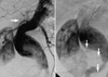
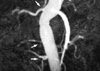
A 73-yr-old male presented to us with a pulsating abdominal mass. Abdominal computerized tomography (CT) scan showed an abdominal aortic aneurysm having crossed ectopia with fusion anomaly of the kidneys. His past medical history was positive for smoking, hypertension, benign prostatic hypertrophy, and parkinsonism. On physical examination, a pulsating non-tender abdominal mass of about 8 cm in diameter was palpable around the umbilicus. Lower extremity pulses were palpable without evidence of aneurysmal dilatation. Laboratory tests, chest radiography, and electrocardiography were normal. The patient underwent a transfemoral aortogram (Fig. 1) to evaluate the renal arterial anatomy. One renal artery from the abdominal aorta for the upper (right) kidney, and two renal arteries from the aneurysm and one renal artery from the left common iliac artery for the lower kidney (crossover left) were seen. One pelvis and one ureter for each kidney were also identified. Operation was conducted. The retroperitoneal cavity was approached transperitoneally. The ureter of the lower (crossover left) kidney crossed the midline at the level of the bifurcation and entered the bladder. To minimize renal ischemic injury, we dissected the lower two renal arteries (one from the aneurysm itself and the other from the left common iliac artery) and anastomosed the lowest renal artery (from the left common iliac artery) to middle renal artery (from the aneurysm itself) in a side-to-end fashion before aneurysm resection. We administered mannitol before the aortic cross clamping. After clamping the abdominal aorta below the level of the right renal artery and bilateral internal and external iliac arteries, the aneurysm was opened and two renal artery orifices were discovered. After the proximal anastomosis of the bifurcated graft, two renal arteries were individually reimplanted and blood flow was resumed to renal arteries. The renal arteries were reperfused within 20 min after the aortic cross clamping. Anastomosis of the two iliac limbs of the bifurcated graft to common iliac arteries was performed without concern of the time. The patient recovered uneventfully and was discharged on postoperative 7th day. Magnetic resonance (MR) angiogram (Fig. 2) was taken two weeks after the operation and showed patent renal arteries and well perfused kidneys.
DISCUSSION
Developmental abnormalities of the kidney are technically challenging to the vascular surgeon at the time of aortic reconstruction, especially during abdominal aortic aneurysm (AAA) surgery due to the presence of aberrant, accessory, or multiple renal arteries, and the risk of unexpected injury to the collecting system (1-4). Crossed renal ectopia is the second most common renal fusion anomaly following the horseshoe kidney with an incidence in the general population of 1 in 1000, and crossover of the left kidney to the right side is the most common form of this variation (1, 2).
Crossed renal ectopia has been further classified depending on the shape assumed by the fused renal masses, but all types are managed similarly during aortic reconstruction (1). Once the diagnosis of renal ectopia or fusion is made, preoperative imaging to identify the vascular and collecting system anatomies is necessary for planning of the optimal strategy for reconstruction of the aorta and renal arteries (1-3). The renal arteries may arise from the aneurysm itself requiring reimplantation or bypass grafting. Renal blood supply must be preserved because all renal and accessory renal arteries are end arteries without communication. The sacrifice of any blood supply to the kidney is disastrous, especially if the other kidney is absent or has impairment of renal function (4). Crossed ectopia is always associated with abnormally located ureters, and the involved ureter or ureters cross the midline at the level of the distal aorta or bifurcation and enter the bladder on the side of embryonic origins (2). Rapid reperfusion of the renal artery is the most critical factor to avoid postoperative renal failure (5). The renal arteries must be identified before the aortic clamping, and either be reimplanted or bypassed within the allowable ischemic time. Otherwise implementation of some protective measures such as cooling of the kidney or temporary shunting is necessary to avoid renal ischemic injury (5). Our method of joining two renal arteries after mobilization before the aortic cross clamping could shorten the renal ischemic time. In conclusion, preoperative imaging to define the arterial and collecting system anatomies and planning of the operation accordingly is essential during AAA repair in a patient with combined renal developmental abnormalities.
 XML Download