

 PDF
PDF ePub
ePub Citation
 Print
Print


INTRODUCTION
Lichen planus pigmentosus (LPP), an uncommon variant of LP, is characterized by mottled or reticulated hyperpigmented, dark brown macules in sun-exposed areas and flexural folds. The histopathologic findings of LPP consist of hyperkeratosis, atrophic epidermis with vacuolar alteration of the basal cell layer, and scarce lymphohistiocyte or lichenoid infiltrates in the dermis with pigmentary incontinence and the presence of melanophages (1). Although there have been a few reports of linear lichen planus, there has been no report of LPP with a linear pattern (2, 3). We describe two patients with LPP who had skin lesions in a linear distribution related to Blaschko's lines on their extremities. This condition should be considered in the differential diagnosis of linear hyperpigmented skin lesions.
CASE REPORT
Patient 1
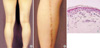
A 23-yr-old Korean woman presented with asymptomatic dark brown macules on the left leg for 2 yr. One or two dark brown macules first appeared and spreaded gradually without any preceding erythematous or scaly skin eruption. There was no history of prolonged sun exposure or trauma on the lesion site. On examination, she had linear streaks of dark brown macules from the left ankle, across the calf and to the thigh, consistent with the pattern of Blaschko's lines (Fig. 1A, B). There was no oral or nail involvement. Skin biopsy showed atrophic epidermis, basal hydropic degeneration with sparse perivascular lymphohistiocytic infiltrates, and numerous melanophages (Fig. 1C). These findings were consistent with LPP.
Patient 2
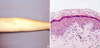
A 16-yr-old Korean woman presented with linear dark brown pigmentation on the left arm for one year. She stated that recurrent asymptomatic erythematous papules first appeared and then the lesion had developed into dark brown macules during 1-2 weeks. Examination showed two linear streaks of dark brown macules from the left dorsum of hand to the upper arm probably related to Blaschko's lines (Fig. 2A). There was no accompanying oral or nail change. Skin biopsy showed orthokeratosis, focal basal liquefaction, a sparse perivascular inflammatory infiltrate, and pigmentary incontinence, confirming the diagnosis of LPP (Fig. 2B).
DISCUSSION
LPP has been described as a condition of unknown etiology which clinically differs from the classical lichen planus by exhibiting dark brown macules and/or papules and a longer clinical course without pruritus or scalp, nail, or mucosal involvement. The LPP is most common on sun-exposed areas such as the face, neck, and flexural folds including the axilla, inguinal, and submammary regions (1). Some authors observed a striking predominance of lesions in an intertriginous location, with most of them in the axillae, thus they proposed the designation LPP-inversus (4). Less common presentations include zosteriform pattern on the trunk (5) and involvement of non sun-exposed areas such as thigh (6). Our patients had lesions on the extremities with a linear pattern. The linearity of the lesions is probably related to Blaschko's lines, which suggests that the predisposition to develop LPP might be determined during embryogenesis.
The differential diagnoses of our cases include lichen striatus (7), linear and whorled nevoid melanosis and incontinentia pigmenti. In particular, because lichen striatus may have post-inflammatory hyperpigmentation (8) and lacks a well-defined histopathological picture (9), it should be differentiated in our patients. Lichen striatus almost always have preceding inflammatory papules or scaly eruption, which last for 4 months to 4 yr (8). There was no previous papule in patient 1 and the short duration of preceding papules (1-2 weeks) favor the LPP in patient 2. In addition, the histological findings in our patients were more consistent with LPP. There were no epidermal changes consisting of spongiosis and exocytosis, and a deeper dermal inflammatory infiltrate around adnexal structures which are features frequently seen in lichen striatus (9).
In summary, LPP can present with a linear pattern and thus should be considered in the differential diagnosis of linear hyperpigmented skin lesions.
 XML Download