

 PDF
PDF ePub
ePub Citation
 Print
Print


Despite there are some debates with catheter closure of the patent foramen ovale (PFO),1) it can be an option in reducing the risk of recurrent paradoxical embolism and incidence of migraines. Although it has been a safe and promising technique with a low incidence of periprocedural complication,2) some of complications during the procedure can be fatal. We found a linear, highly mobile lesion attached to the PFO closure device during the procedure. What did happen during the procedure?
A 48-year-old woman visited the emergency room with an acute onset of dysarthria. She was diagnosed with a cerebral infarction of the superior branch of the right middle cerebral artery. The patient had a history of transient ischemic attack about 4 years ago. At the time, her transesophageal echocardiography revealed the presence of about a 1 mm sized PFO with a significant right to left shunt through it after having the Valsalva maneuver performed. She discontinued her medication after one month and did not visit the out-patient clinic. Because of the recurrent embolic events, the attending neurologist and consultant cardiologist decided to occlude the PFO with the Amplatzer® PFO occlude device (AGA medical Co., Minneapolis, MN, USA).
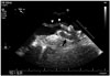
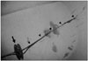
The intervention was performed under local anesthesia with fluoroscopic guidance. After a venous puncture of the right femoral vein, the PFO was crossed under fluoroscopic guidance in the anteroposterior view by a standard length normal 0.035-inch guidewire. Initially, a J-tip was used to ensure passage through the hole. The interventionist tried to insert a 23-mm device through the J-tip. However, the procedure was not smooth and repeated attempts were performed. The device was finally inserted after several attempts. The follow-up echocardiography showed a freely moving linear mass lesion on the tip of the left atrial disc. The transesophageal echocardiography demonstrated about a 13 cm sized linear mass attached to the tip of the left atrial disk (Fig. 1, Supplementary movie 1). The highly mobile mass moved to-and-fro through the mitral valve. To reduce further catastrophic embolic events, the patient underwent open heart surgery to remove the device with the mass and occlude the PFO. We found with about a 20 cm sized linear fresh thrombus attached to the tip of the left atrial disc (Fig. 2). Fortunately, she was discharged without further complications after the surgery. Meticulous handling is necessary to prevent this injurious complication during procedure.
 XML Download