

 PDF
PDF ePub
ePub Citation
 Print
Print


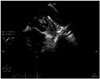
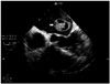
Left atrial (LA) thrombus is known as a common complication of rheumatic mitral stenosis. It is more common in the presence of atrial fibrillation (AF). Incidence of LA clot formation in sinus rhythm is 2.4-13.5%1)2) and the incidence is as high as 33% in patients with mitral stenosis in AF. Because transthoracic echocardiography may not detect the left atrial appendage (LAA) thrombus, transesophageal echocardiography (TEE) remains the gold standard for identifying the LAA thrombus. We report a case of LAA thrombus mimicking like a hydatid cyst. A 38 years old male with severe mitral stenosis due to rheumatic heart disease presented with progressive worsening of dyspnea (New York Heart Association class II-III) over a period of two years. The electrocardiogram revealed sinus rhythm with evidence of LA enlargement. There was no past history of AF and he was not on any anticoagulants. The transthoracic echocardiogram revealed thickened mitral leaflets with mitral orifice area of 1.0 cm2 and mean mitral valve gradient of 18 mm Hg. The LA was enlarged and there was grade IV spontaneous echo contrast in LA. On TEE, there was large cystic mobile formation noted in the LA, originating from LAA and protruding into LA body. Nodular structures were visible within the cystic formation (Fig. 1, Supplementary movie 1). In four-chamber view at 0°, it was appearing like a blooming flower (Fig. 2, Supplementary movie 2). Thrombus is identified by the homogenous echo texture and visualised in more than one plane. It can appear in various forms and shapes depending on the organisation of clot.
TEE is a useful tool to detect LA thrombus in patients with mitral stenosis. The clot in LA can occur in various forms from cystic lesions to mass like appearences. One should look for LAA clot in cases of rheumatic mitral stenosis before subjecting for balloon mitral valvotomy whether patients are in AF or sinus rhythm. In the presence of high grade of spontanous echo contrast, LA clot should be always suspected.3)
 XML Download