Abstract
A 55-year-old male presented with stroke. Transesophageal echocardiogram and cardiac computed tomography revealed an unrecognized congenital malformation of the anterior mitral leaflet associated with anomalous left coronary circumflex artery, arising from the right coronary artery, diagnosed first by echocardiogram. This case represents a unique unforeseen mitral valve anomaly that might be considered as potential cardiac source of embolism. This finding broadens the spectrum of known mitral valve anomalies.
Congenital mitral valve anomalies are very rare clinical entities involving one or more components of the mitral valve apparatus. The majority of these abnormalities are causing mitral valve insufficiency and commonly presenting in the early part of life. Common clinical presentations include left ventricular outflow tract obstruction, valvular insufficiency and heart failure. We report a rare and unique case of congenital mitral valve anomaly associated with anomalous coronary artery in a middle age man who presented with stroke.
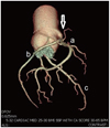
A 55-year-old male, smoker and previously healthy, presented to our emergency department for abrupt onset of difficulty reading and right sided weakness, signs and symptoms suggestive of a new onset stroke. Stat computed tomography (CT) and computed tomography angiogram (CTA) of the brain revealed no abnormality. Magnetic resonance imaging of the brain showed acute infarction of the left posterior insular cortex and left parietal subcortical area. The patient was started on aspirin, atorvastatin and clopidogrel. He underwent a routine transthoracic echocardiogram (TTE) as to rule out any possible cardiac source of embolism. TTE revealed an abnormally thickened anterior mitral leaflet associated with mild eccentric mitral regurgitation (MR). Transesophageal echocardiogram (TEE) was performed for further assessment. TEE revealed some form of redundant tissue along the anterior mitral leaflet and the base of the anterior leaflet in the left ventricular outflow tract (LVOT). It is thin and supple, the appearance was neither consistent with a fibroelastoma, nor a tumor, an abscess, a true aneurysm of the sinus of Valsalva nor perimembranous septum. The anterior mitral leaflet is itself thickened and prolapses, which might represent myxomatous disease. A mild eccentric posteriorly directed MR jet was noted, likely originating from the mild prolapse of the anterior leaflet. At the very distal end of the anterior leaflet, there is an echo bright, independently mobile soft tissue entity, 2-3 mm in size. The left circumflex coronary artery (LCX) appears to have both an anomalous origin (very low in the left sinus of Valsalva immediately above the annulus, in closer association to the mitral valve than usual). The ostium of the right coronary artery appears larger than the left coronary artery (Fig. 1, 2, and 3, Supplementary movie 1, 2, and 3). Based on these findings, an electrocardiogram-gated cardiac CT scan was suggested. Coronary CTA revealed; anomalous LCX originating from the proximal right coronary artery off the right sinus of Valsalva with a retro-aortic benign course. The mitral valve was described as; thickened anterior mitral valve leaflet with a band like attachment to the membranous inter-ventricular septum, the portion of the band closer to the anterior leaflet forms a thin curvilinear band (Fig. 4 and 5). This abnormal appearance could represent a congenital anomaly, myxomatous changes are still probable although may not explain the atypical attachment site. The patient had no fever, blood cultures were negative and all other laboratory investigations were within normal limits. The patient remained in hospital for one week and discharged later with a minor neurological deficit. In the absence of obstruction of the LVOT, the patient is being followed up without surgical intervention.
Congenital malformations of the mitral valve are very rare clinical entity. Most commonly, mitral valve prolapse due to myxomatous degenerative disease and connective tissue diseases, congenital mitral stenosis, accessory valvular tissue, isolated cleft mitral valve, double orifice and Ebstein-like malformation were also reported. Supra-mitral ring is one of the components of Shone's complex. In addition, there are also anomalies of the tensor apparatus i.e., archade or hammock valve, straddling mitral valve with abnormal attachment of mitral valve chordae to both ventricles and parachute mitral valve characterized by unifocal attachment to a single or fused papillary muscle.1,2,3) Blood cyst of the sub-valvular apparatus of the mitral valve are commonly reported as postmortem findings in infants and they regress spontaneously by age of 6 months, few cases in adults resulting in embolisation and valvular dysfunction have been reported.4)
Accessory mitral valve tissue is a rare congenital cardiac anomaly, commonly associated with other congenital heart diseases, such as ventricular septal defects and transposition of the great arteries. Prifti et al.5) have classified accessory mitral valve tissue as Type I-Fixed type (A: nodular, B: membranous), Type II-Mobile type (A: pedunculated, B: leaflet-like). Type IIB is divided into 1) rudimentary chordae and 2) developed chordae. In this case, the appearance might fit into Type IIb-Mobile type (leaflet like). However, they further described attachment of the mitral accessory tissue into six different locations in the left ventricle through the accessory tissue's chordae. In our case, the accessory tissue was attached to the interventricular septum directly, not through a chordae, confirmed by echocardiogram and CT scan.
In the majority of cases of accessory mitral valve tissue, presentations were left ventricular outflow tract obstruction, significant valvular insufficiency, palpitation and heart failure. The LVOT obstruction is the result of systolic ballooning into the LVOT producing a mass effect and the continuing turbulence producing this effect, results in a fibrous tissue reaction with permanent deposits of fibrous tissue and progressive LVOT obstruction.6,7,8) In this case, there was no LVOT obstruction and the presentation was stroke. Mycotic aneurysm of infective endocarditis might give a similar appearance. However, infective endocarditis was excluded in this case on the basis of negative blood culture and absence of other criteria suggesting endocarditis. We reviewed all echocardiographic features of reported mitral valve anomalies, and none have shown similar appearance to the anomalous mitral valve presented in this case.
Echocardiography provides adequate imaging of the origin and proximal course of coronary arteries.9) The transesophageal echocardiogram revealed an anomalous course of the LCX, later confirmed by CTA. The associated coronary anomaly is relatively frequent and might be an unrelated coexistence.
Pathological diagnosis of this uncommon mitral valve anomaly is unavailable, which might be considered as a limitation. However, given the clinical presentation of stroke, there is an ongoing debate, as to consider this anomaly being a definite cardiac source of embolism. In addition, there was no LVOT obstruction and the final decision was to treat the patient conservatively with regular follow-up, which was also the patient's preference.
This case highlights an under-recognized anomaly of the mitral valve associated with anomalous coronary arteries. We advise regular clinical and echocardiographic evaluation in this situation to identify any progression. In future, such anomaly might be considered as potential source of embolism and surgical removal might be considered as a reasonable indication.
Figures and Tables
Fig. 1
Transesophageal echocardiogram. Midesophageal long-axis view. The abnormal accessory tissue attached to the basal portion of the anterior mitral leaflet (arrow A). Origin of the left coronary artery (note the abnormal low origin in the sinus of Valsalva) (arrow B). Origin of the right coronary artery, note the larger vessel caliber (arrow C). Ao: aorta, LA: left atrium, LV: left ventricle, RV: right ventricle.

Fig. 2
Transesophageal echocardiogram. Midesophageal 4-chamber view at 0 degrees. The course of the left circumflex coronary artery (arrow A). The abnormal accessory tissue attached to the basal portion of the anterior mitral leaflet (arrow B). LV: left ventricle, RA: right atrium, RV: right ventricle.

Fig. 3
Transesophageal echocardiogram. Midesophageal 4-chamber view at 40 degrees with colour Doppler. A: In systole, there as a mild eccentric mitral regurgitation jet (arrow). B: In diastole, small jet of flow acceleration at the site of the accessory mitral tissue (arrow).

References
1. Séguéla PE, Houyel L, Acar P. Congenital malformations of the mitral valve. Arch Cardiovasc Dis. 2011; 104:465–479.

2. Banerjee A, Kohl T, Silverman NH. Echocardiographic evaluation of congenital mitral valve anomalies in children. Am J Cardiol. 1995; 76:1284–1291.

3. Bär H, Siegmund A, Wolf D, Hardt S, Katus HA, Mereles D. Prevalence of asymptomatic mitral valve malformations. Clin Res Cardiol. 2009; 98:305–309.

4. Park MH, Jung SY, Youn HJ, Jin JY, Lee JH, Jung HO. Blood cyst of subvalvular apparatus of the mitral valve in an adult. J Cardiovasc Ultrasound. 2012; 20:146–149.

5. Prifti E, Frati G, Bonacchi M, Vanini V, Chauvaud S. Accessory mitral valve tissue causing left ventricular outflow tract obstruction: case reports and literature review. J Heart Valve Dis. 2001; 10:774–778.
6. Cil H, Atilgan ZA, Islamoglu Y, Yavuz C, Tekbas EÖ. Asymptomatic and isolated accessory mitral valve tissue in adult population: three case reports and review of the literature. Eur Rev Med Pharmacol Sci. 2012; 16:Suppl 4. 74–77.
7. Singh B, Srinivasa KH, Surangi MJ, Rangan K, Manjunath CN. Anomalous mitral arcade variant with accessory mitral leaflet and chordae presenting for the first time with acute decompensated heart failure in an adult. Echocardiography. 2013; 30:E202–E205.


 PDF
PDF ePub
ePub Citation
 Print
Print


 XML Download