

 PDF
PDF ePub
ePub Citation
 Print
Print


Introduction
Primary tumours of heart are rare across all age groups with a reported prevalence of 0.001 to 0.03% in autopsy series.1) About 70% of all primary cardiac tumours are regarded as benign neoplasms. Even though histologically benign they may have potentially devastating effect by virtue of their anatomic location and resultant arrhythmias with fatal consequences. We report one such case.
Case
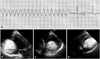
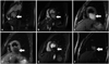
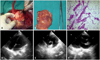
A 23-year-old male was referred to us for evaluation of three episodes of syncope in the preceding week. He was asymptomatic prior to the onset of presenting symptoms. Physical examination was unremarkable. Routine investigations like haemogram, random blood sugar, serum creatinine and serum electrolytes were within normal limits. There was no appreciable change in cardiac silhouette on X-ray chest. In-hospital electrocardiogram obtained at the time of syncope revealed wide complex regular tachycardia with rate of 250 beats/min without clear definition of QRS and T waves suggestive of ventricular flutter which spontaneously terminated (Fig. 1A). The RBBB morphology, positive QRS complex in lead aVL with negative complex in lead I and inferior leads indicated origin form posterior wall of left ventricle. Transthoracic echocardiogram showed a smooth surfaced hyper-echoic mass (8 × 4.8 cm) in the mid and apical region of the left ventricular cavity arising from its postero-inferior wall with normal pericardium (Fig. 1B, C, and D, Supplementary movie 1). Differential diagnosis of cardiac tumors like myxoma, lipoma, hamartoma and metastasis were considered. Cardiac magnetic resonance imaging evaluation of the mass, on T1-weighted (Fig. 2A and B) and double inversion recovery image (Fig. 2C), showed a hyper intense mass which was suppressed in triple inversion (Fig. 2D) and short tau inversion recovery (STIR) sequence (Fig. 2E) with areas of delayed contrast enhancement suggestive of fibro fatty tissue (Fig. 2F). Surgery was subsequently performed via median sternotomy. Left ventricular cavity was approached through anterior wall, close to the apex. Debulking of yellowish white glistening encapsulated mass of size 8.5 × 5.5 × 4.5 cm from left ventricular myocardium was done (Fig. 3A and B). Residual space was obliterated with pericardial pledgets sutures. Histopathology of the mass revealed mature fat cells with ocpcasional fibrous connective tissue consistent with the diagnosis of fibrolipoma (Fig. 3C). Transthoracic echocardiogram in the post operative period showed complete clearance of tumor in left ventricle and good systolic function (Fig. 3D, E, and F, Supplementary movie 2). Since the resection was visually complete and patient did not experience any arrhythmic event during hospital stay, treatment with oral antiarrhythmics was discontinued. Holter monitoring after one month of surgery showed no tracings of ventricular arrhythmia. He is asymptomatic at 6 months follow-up.
Discussion
Cardiac lipoma is rare, accounting for only 0.5-3% of excised heart tumours. The histopathological variant, fibrolipoma, is even rarer and is inadequately described in the literature. It can occur sporadically at all ages with equal sex predilection, most being asymptomatic and incidental finding.1) Symptoms are directed based on tumor size and its location on atrial, ventricular surface or valvular surface.2)3) Intra-myocardial fibrolipoma may interfere with electrical conduction in the heart causing arrhythmias. Mechanisms include compression of the His bundle resulting in conduction block, creation of re-entry pathways producing ventricular tachycardia, regions of asynchronous refractoriness within the tumour mass producing electrical instability, bradyarrhythmias and ventricular fibrillation.4)5) The echocardiographic appearance of cardiac fibrolipoma is nonspecific and varies with the location of tumor. Lipomas situated in the pericardial space have variable echogenecity but are often hypoechogenic, while on the contrary, intracavitary lipomas are typically homogeneous and echogenic. The reason for this difference is unknown. Fibrolipoma, owing to its fibrous content interspersed between fat cells, is typically heterogenous. At echocardiography, intracavitary lipomas are usually circumscribed but cannot be differentiated from other circumscribed cardiac masses. Differential diagnoses of such cardiac masses are myxomas, lipoma, fibrolipoma, hamartoma, mesenchymoma. A cardiac computed tomography (CT) and magnetic resonance imaging (MRI) allows for very specific identification of fat and therefore can be used for better tissue characterization. In cardiac CT, lipomas appear as homogeneous, low-attenuation masses with no contrast enhancement either in a cardiac chamber, walls or in the pericardial space.6) The MRI of fibrolipoma is characterized by homogeneous bright signal intensity on T1-weighted and double inversion recovery images that decreases on T2-weighted images and is suppressed on triple inversion recovery sequences. However few areas show hypointensity in all the sequences. First pass perfusion weighted image shows no change in the signal intensity of the tumor on contrast administration but the areas with hypointense signal shows delayed contrast enhancement suggestive of fibrolipoma. A decrease in signal intensity using a fat presaturation sequences and delayed contrast enhancement verifies the diagnosis. Histopathologically, cardiac fibrolipoma is composed of circumscribed masses of mature adipocytes with occasional fibrous connective tissue. Symptomatic cardiac fibrolipomas after surgical excision have excellent prognosis.7)
Majority of symptomatic cardiac lipomas occurs in pediatric population and usually present with arrhythmias. Fibrolipoma involving the left ventricle in adult is a rare occurrence. Fibrolipoma should be included in the differential diagnosis of cardiac mass in adult population. Structural heart disease should be ruled out by any imaging modality in patients with arrhythmias.

 XML Download