

 PDF
PDF ePub
ePub Citation
 Print
Print


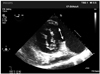
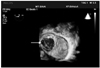
A 50-year-old woman presented to the operating room for repair of a left ventricular pseudoaneurysm. She had a history of cardiac sarcoidosis which had manifested as dilated cardiomyopathy and recurrent ventricular tachycardia, which was treated with an endocardial/epicardial ablation six months ago. Transesophageal echocardiography at the transgastric basal short axis view demonstrated a 4 cm rupture of the basal inferolateral wall of the left ventricle close to the base of the posterior mitral valve annulus giving rise to a large (9.5 × 7 cm) pseudoaneurysm (Fig. 1). Three dimensional reconstruction (Fig. 2) as seen from within the pseudoaneurysm looking into the left ventricle shows the extent of the rupture. The mitral valve apparatus can be seen through the defect. The patient underwent a patch closure of the defect.
The fundamental features that differentiate true aneurysms from false aneurysms are the presence of a continuous surrounding myocardial wall found in true aneurysms and the presence of a pericardial wall usually with mural thrombus found in false aneurysms.1) Due to its lack of structural myocardial support, a left ventricular pseudoaneurysm carries a significant risk of expansion and fatal rupture and therefore requires an urgent surgical intervention.2)
 XML Download