

 PDF
PDF ePub
ePub Citation
 Print
Print


Introduction
In developed societies, the most common etiology of severe mitral regurgitation (MR) is degenerative valve disease, resulting in mitral valve prolapse (MVP) or flail mitral leaflet (FML). Although corrective mitral valve (MV) surgery is the only efficient treatment, the optimal timing of intervention in the individual patient is often a challenging, multifaceted decision which is highly contingent on local resources and expertise. Accurate echocardiographic evaluation and early referral for surgical intervention can achieve excellent long-term outcomes but strategies entailing careful patient surveillance and risk stratification may not be inferior. This review examines the current perspective on surgical timing in degenerative MR, with a focus on the role of cardiac imaging and in particular, echocardiography.
Pathophysiology and "Natural History"
In chronic MR, increasing preload imposes a volume overload on the left ventricle (LV). The LV dilates to delivers a normal forward cardiac output despite regurgitation into the left atrium (LA). This LV remodeling into eccentric hypertrophy occurs through rearrangement of myocardial fibers and addition of new sarcomeres in series.1) Although LV dilatation should result in an increase in afterload, this is reduced by ejection of much of its volume into the low-impedance LA. Over time, the hemodynamic burden of volume overload eventuates in LV dysfunction, impaired ejection and increased filling pressure.2)
The clinical evolution of severe MR parallels these pathophysiologic changes. In the compensatory phase, the patient may be asymptomatic or have minimal symptoms for several years. Eventually, symptoms of pulmonary congestion, reduced cardiac output and pulmonary hypertension supervene, sometimes abruptly with the onset of atrial fibrillation (AF).3) The clinical course is not always predictable - LV dysfunction can occur in the absence of symptoms,4) and sudden death, while uncommon, is a recognized sequela, even in subjects with no or minimal symptoms.5) In the era of preemptive cardiac surgery, the true natural history of MR is difficult if not impossible to appraise. In a retrospective study of 229 patients with flail leaflet, 71% of whom had New York Heart Association (NYHA) class I-II symptoms at baseline, the reported rates of heart failure (HF), AF, and death or surgery at 10 years were 63%, 30%, and 90%, respectively.6) Besides older age, presence of symptoms and low LV ejection fraction (LVEF), surgery, performed at any time during follow-up, was an independent determinant of mortality.
Management Strategies and Outcomes
It is universally accepted that MV surgery is the only effective treatment that can favorably reverse the LV response to volume overload. Traditionally, referral for surgery was triggered by the occurrence of symptoms or LV dysfunction. If not operated upon in timely fashion, patients with FML and NYHA III-IV symptoms have a 34% annual mortality despite initial functional improvement with medical therapy.6) Hence, for symptomatic patients with chronic severe MR, the indication for surgery is not controversial. The 2008 American Heart Association-American College of Cardiology7) and 2007 European Society of Cardiology8) valvular heart disease guidelines recommend surgery in these subjects unless there is severe LV dysfunction, defined as LVEF < 30% and LV end-systolic diameter (LVESD) > 55 mm (see also later comments on the updated European guidelines). For severely symptomatic subjects with even more severe LV impairment, valve repair if feasible can be considered. In general, MV repair is preferred to replacement in the majority of patients who should be referred to surgeons skilled in repair techniques, and optimally to a recognized "reference" valve centre when there is multisegmental or Barlow's disease.9) Advantages of repair include avoidance of long-term anticoagulation, and preservation of more physiological MV function, including mitral annulus-papillary muscle continuity, which translates into better LV function.10) Outcomes are also superior after valve repair, with an operative mortality of 1% as opposed to nearly 4% in a contemporary series,11) and long-term survival better.12)
For asymptomatic subjects, the guidelines recommend valve surgery for patients with LVEF < 60% or LVESD ≥ 40 mm (> 45 mm by European guidelines).7)8) Additionally, surgery is recommended even in the absence of symptoms or LV dysfunction when AF ensues or when pulmonary pressures are elevated to ≥ 50 mmHg at rest or ≥ 60 mmHg during exercise. For asymptomatic subjects without any of these risk factors, the timing of surgical intervention has been the subject of considerable debate. The Dutch Asymptomatic Mitral Regurgitation trial aims to directly address this issue by randomizing 250 asymptomatic patients with severe organic MR and preserved LV function to MV surgery or medical observation, and comparing outcomes over 5 years.13)
In the absence of randomized trial data, prospective and retrospective studies in asymptomatic patient populations can help inform clinical management. Enriquez-Sarano et al.14) prospectively followed 456 asymptomatic patients with variable degrees of MR and LVEF > 50%. Of these, 198 had severe MR defined by an effective regurgitant orifice area (EROA) ≥ 40 mm2; their LVEF was 70 ± 8%. At 5 years, these patients had a high cardiac event (i.e., cardiac death, HF and new-onset AF) rate of 62 ± 8%, and survival rate of only 58% compared to 78% for a matched control population. Surgery, ultimately performed in 232 patients, was independently associated with improved survival. Although this Mayo Clinic study highlighted the poorer outcomes in conservatively managed patients with severe MR by quantitation, the propriety of intervention was determined independently by the primary physician rather than by formalized, uniform criteria.
Using a protocolized guidelines-driven approach to management, Rosenhek et al.15) prospectively followed 132 asymptomatic patients with severe degenerative MR with preserved LV function and in sinus rhythm. Patients underwent serial clinical and echocardiographic examinations by the same group of physicians at 3-6 monthly intervals and were referred for surgery only when standard guidelines criteria were fulfilled. During follow-up, 38 patients developed indications for surgery - 24 had symptoms, 9 asymptomatic LV dysfunction or enlargement, and 5 new onset pulmonary hypertension or AF. Overall survival was not significantly inferior at 8 years compared with a control population. While this "watchful waiting" approach does appear to justify consensus recommendations, the conservative strategy was associated with a nearly 50% rate of progression to surgical indications within 5 years and notable mortality (8 deaths) during follow-up. Postoperative LVEF was also reduced in 4/35 (11%) of patients who had surgery. Thus, while conservative surveillance appears to be a "safe" initial approach, this should be conducted with vigilance, preferably in a dedicated valve clinic setting.15)16) This strategy may be germane to certain subgroups: the very elderly, those in whom repairability of the MV is in considerable doubt, and patients with comorbidities which increase the risk of surgery as well as prosthetic valve complications.
More recently, Kang et al.4) reported the long-term results of a prospectively conducted Korean registry of 447 asymptomatic patients with severe degenerative MR and preserved LVEF - 161 patients underwent early surgery and 286 were managed conservatively. Over a 9 year period of follow-up, there were only 2 cardiac events (both repeat surgeries) in the early surgery group compared to 35 (12 cardiac deaths, 22 hospitalizations for HF, and 1 repeat surgery) in conservatively managed patients, yielding 99% and 85% cardiac event-free survival rates, respectively. Propensity analysis of 127 surgically treated patients matched for baseline characteristics with 127 conservatively managed subjects showed significantly better outcomes in the former. Likewise, Montant et al.17) used propensity analysis to compare outcomes in 125 patients who had early MV repair and 67 patients managed conservatively over a median of 8.5 years. They estimated 10-year hazard ratios for overall and cardiac mortality, and cardiovascular events, of respectively, 5.21, 4.83, and 4.40 in conservatively treated patients. While these studies ostensibly support prompt surgical intervention in asymptomatic severe MR, they are again non-randomized and unaccounted confounders could have biased the results. In the study of Kang et al.4) for example, the very high proportion (94%) of conservatively managed patients who were symptomatic at eventual surgery suggests undue delay in crossover, possibly in part because of perceived more difficult repair.18)
Results of Mitral Valve Repair
Notwithstanding the limitations of non-randomized studies, early surgery for severe organic MR, in particular MV repair, is increasingly encouraged. In the Society of Thoracic Surgeons Adult Cardiac Surgery Database, MV repair rates for isolated MR rose from 51 to 69% between January 2000 and December 2007.19) Operative mortality was significantly lower for repair compared to MV replacement even after risk-adjustment (odds ratio, 0.52), and among asymptomatic patients, only 0.6%. The low mortality rate aside, MV repair does entail the risk of unintended valve replacement, with attendant risks of anticoagulation if a mechanical valve is implanted, as well as future reoperation, especially if there is residual MR. In one series, moderate to severe MR recurred at a linearized 3.7% per annum, a rate only partially attributable to inadequate surgical technique.20) Even in accomplished surgical centres, the reoperation rate is 5-10% at 10 years, more frequently after anterior leaflet repair.21) Recently, Castillo et al.9) reported a 97% freedom from reoperation rate at 7 years among 744 patients with degenerative MV disease in whom a repair strategy was applied systematically in all cases. However, the true durability of MV repair could not be ascertained as only 70% of patients had late follow-up echocardiograms. These impressive results were attained by a single surgical team and cannot be generalized since the likelihood of successful repair is dependent on surgical volume22) and appears surgeon-specific.23) Clearly, patients with technically challenging pathology such as Barlow disease with multi-segmental leaflet billowing are best served by a "reference" valve surgeon.24) Equally, the enthusiasm to recommend preemptive surgery to asymptomatic patients should be tempered by a realistic assessment of locally available surgical expertise. In the absence of "high-risk" features, intervention should logically be considered only if the expected success rate of MV repair within the specific healthcare setting exceeds 90%.7)
Why Risk Stratify the Asymptomatic Patient?
Because the outcomes of MV surgery are not optimal in all cases, all authoritative guidelines include clinically accessible measures of LV remodeling and "function" to identify asymptomatic patients at higher risk. These criteria, elaborated upon earlier, were largely derived from studies on preoperative determinants of outcome after surgery which do not necessarily represent optimal thresholds for intervention. For instance, in the study of Matsumura et al.,25) 6-7% of patients developed postoperative LV dysfunction despite having an LVESD < 40 mm at outset. Long-term outcomes data from a large Mayo Clinic cohort of 1063 patients who had MV repair or replacement indicate a greater likelihood of optimal LVEF recovery i.e. > 60% if preoperative LVEF was > 65% (hazard ratio, 1.7) or LVESD < 36 mm (hazard ratio, 2.0).26) Since severe MR is a progressive condition which entails the risk of LV dysfunction and frequent need for intervention,15)27) additional means of risk stratification are desirable. Ideally, these should help ascertain the ideal timing of intervention to preserve LV function and not simply avert a poor outcome. In this regard, symptoms are unreliable - they are ambiguous in the elderly and deconditioned, often minimized by voluntary limitation of physical activity, and can be absent in the face of objective LV dysfunction.4) Risk stratification may also provide reassurance that intervention can be safely delayed in asymptomatic patients who remain undecided about operation, in those whose interests are not best served by immediate surgery or have other extenuating circumstances necessitating deferral of surgery.
Role of Rest Echocardiography
Risk stratification in patients with organic MR is heavily reliant on echocardiography which plays a pivotal role in evaluating the effects of volume overload.7)22) Apart from determination of LV dimensions, volumes and function, direct quantitation of MR is performed routinely in many echocardiographic laboratories, often using the proximal isovelocity surface area (PISA) technique. Since, among asymptomatic patients, an EROA > 40 mm2 portends significantly poorer outcomes in the long term, it may be justified as suggested by the Mayo Clinic group to consider prompt corrective surgery in these individuals.14) Adoption of such a policy does however mandate highly accurate quantitation of MR. While exponents of the PISA method vouch for its accuracy and reproducibility,28)29) others have found only modest reliability and significant interobserver variability. In one study involving 11 academic centres, the rate of "substantial agreement" among echocardiologists for distinction of severe from non-severe MR using EROA was only 38%.22) Other limitations of the two-dimensional color Doppler PISA technique, including geometric assumptions of the flow convergence zone, have become apparent from three-dimensional echocardiographic and cardiac magnetic resonance studies.30)31) The validity of using the standard PISA technique to quantitate predominantly mid-to-late systolic regurgitation characteristic of MVP is also in doubt.
Given these pitfalls and the absence of an unimpeachable reference technique, assessment of MR severity should integrate quantitative and qualitative data, including vena contracta width, pulmonary vein systolic flow reversal, and presence of FML. LA size and pulmonary artery systolic pressure (PASP) are also relevant information. The importance of the LA in the natural history of MR has been under-appreciated and until recently, not figured in guidelines. This is a surprising oversight since LA enlargement reflects, among other factors, the regurgitated volume and LV dysfunction and therefore the chronic hemodynamic burden of organic MR. In an earlier study, Reed et al.32) showed that the "LA index", determined by multiplying longitudinal and transverse LA dimensions from the apical 4-chamber view, independently predicted cardiac death among 176 patients who underwent MV replacement. More recently, the Mitral Regurgitation International Database investigators found an increasing mortality hazard for conservatively managed patients with FML and anteroposterior LA dimension ≥ 55 mm.33) This same group has also shown that pulmonary hypertension, defined as resting PASP > 50 mmHg, significantly increases total and cardiac mortality under medical management, a risk ameliorated by but not completely abolished by MV surgery.34)
Stress Echocardiography
Isotonic exercise produces a dramatic increase in myocardial contractility in normal subjects,35) and may be a useful means of detecting latent LV dysfunction in asymptomatic patients with severe MR and "preserved" resting LVEF. Stress echocardiography can evaluate contractile reserve (CR) and other metrics such as functional capacity and exercise-induced pulmonary hypertension which may have prognostic value. In a study of 74 patients with organic MR who had echocardiography before and after treadmill or upright bicycle exercise, Leung et al.36) showed that post-exercise LV end-systolic volume index > 25 mL/m2 had superior sensitivity and specificity (both 83%) for predicting LV dysfunction after MV repair to all measures of resting LV performance. Lee et al.37) found that surgically treated MR patients with CR (defined by augmentation of LVEF by at least 4% during exercise) had better preserved LVEF out to 3 years, while persistent postoperative LV dysfunction (i.e., LVEF < 50%) occurred only in those without CR. Among medically followed subjects, LVEF remained stable in those with CR but progressively deteriorated when CR was absent. CR was not only an independent predictor of follow up LVEF but also prognostic - at 38 months, postoperative cardiac events occurred in 50% of patients without CR and in no patient with CR. Exercise echocardiography can also unmask pulmonary hypertension in asymptomatic degenerative. Exercise-induced pulmonary hypertension (PASP > 60 mmHg) is associated with markedly lower 2-year symptom-free survival, and appears superior to resting pulmonary hypertension for predicting the occurrence of symptoms.38)
Cardiopulmonary exercise testing may also have a role in risk stratification. Messika-Zeitoun et al.39) showed that reduced functional capacity (defined as peak oxygen consumption < 85% of expected) was associated with a significantly higher rate of clinical events (death, HF or new AF) or the composite of these events and MV surgery at 3 years. This risk persisted even after adjusting for age and EROA (risk ratios, 1.8 and 1.5, respectively).
Tissue Doppler and Myocardial Deformation Imaging
In chronic organic MR, incipient LV dysfunction may be detected by assessing LV long axis function which appears to be deranged earlier than radial shortening. Haluska et al.40) were one of the first to investigate tissue Doppler imaging (TDI) in the setting of asymptomatic or minimally symptomatic MR. They showed that peak annular systolic velocity (Sm) was the only resting echocardiographic parameter that could discriminate patients with and without CR; resting LVEF, LV volumes or sphericity index did not. The differences in peak Sm were even greater after stress. Being a marker of CR, Sm may potentially predict postoperative LV function. In one study, Sm ≤ 10 cm/s was independently associated with a > 10% postoperative reduction in LVEF.41)
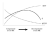
Compared to TDI, myocardial strain imaging is less influenced by cardiac translation and tethering. Whether early LV decompensation is heralded by a decrease in deformation indices has been the subject of recent investigation. Among 27 asymptomatic MVP patients with "moderate to severe" MR (mean regurgitant fraction, 59%), Borg et al.42) found instead increased Doppler-derived systolic strain and strain rate compared to controls. Contrasting findings were reported by Marciniak et al.43) who studied 54 asymptomatic or minimally symptomatic patients with variable degrees of organic MR and 23 healthy subjects, and observed a moderate negative correlation between end-systolic strain and peak systolic strain rate. The correlation improved after correcting these regional deformation indices for LV end-diastolic volume. Mathematical modeling indicated a complex dependency of myocardial deformation on LV size, stroke volume and LV contractility, and not directly on MR severity per se. Differences in patient characteristics may account for the discrepant strain imaging findings in these 2 studies. The subjects studied by Borg et al.42) had normal or supranormal LVEF (mean, 68%) and appear to be in the compensated phase of volume overload while the severe MR patients of Marciniak et al.43) had a mean LVEF of 61 ± 12%, and therefore likely, in some cases, to have incipient or even frank LV dysfunction. Collectively, the findings indicate that ejection phase strain indices are load dependent, as is increasingly recognized.44) "Pseudonormalization" of myocardial strain in chronic MR, whereby fiber shortening is enhanced in the compensated state but later decreases with disease progression (Fig. 1), suggests that normalization for preload or LV geometry is required to appropriately interpret this parameter.45) The biphasic behavior of myocardial strain may also account in part for conflicting reports on the value of preoperative speckle-tracking strain imaging in predicting postoperative LV dysfunction.46)47)
Natriuretic Peptides
As in a myriad of other cardiac conditions, B-type natriuretic peptide (BNP) may have a role in risk stratification of MR. Sutton et al.48) were the first to report an association between BNP (as well as aminoterminal pro-BNP and atrial natriuretic peptide) and cardiac-related symptoms in patients with organic MR. Detaint et al.49) found BNP to be an independent predictor of mortality and/or HF in 124 patients with variable degrees of functional limitation and MR severity. In a prospectively followed cohort of 269 asymptomatic, severe MR patients with LVEF > 60%, Pizarro et al.50) showed that a plasma BNP level > 105 pg/mL was the strongest independent predictor of an unfavorable composite outcome of HF, LV dysfunction or death at follow-up. The high negative predictive value (94-96%) of normal or lower BNP levels may be particularly helpful in identifying patients at low risk for cardiac events. Serial measurements of BNP may further aid risk stratification of these patients.50)51)
2012 European Guidelines on Surgery for Organic Mitral Regurgitation
The recently updated guidelines on management of valvular heart disease issued by the Joint Task Force of the European Society of Cardiology and European Association for Cardio-Thoracic Surgery considers some of the recent research into degenerative MR outlined in this review.52) Recommendations for intervention in asymptomatic patients are summarized in Table 1. There is consensus that surgery is beneficial when there is evidence of LV dysfunction but when LV function is "preserved", repair "should be" or "may be" considered if additional risk factors are present. Of note, the asymptomatic patient with "preserved" LV function, a high likelihood of lasting repair and estimated low surgical risk, is considered a candidate for surgery if two additional criteria are satisfied i.e. presence of FML and LVESD ≥ 40 mm. This is somewhat more conservative than the 2008 American College of Cardiology-American Heart Association guidelines, but does safeguard against premature surgery for MVP with less than severe MR. LA size and exercise-induced pulmonary hypertension are now accounted for, albeit as a weak indication for intervention. The 2012 statement also acknowledges a prognostic role for BNP in MR but does not factor this biomarker in the consideration on surgical timing. Notably, all recommendations for repair in the asymptomatic MR population were under-girded by low evidence levels, emphasizing the need to expand the clinical evidence base.
Conclusions
The optimal timing of surgical intervention is a crucial landmark in the management of degenerative MR, a progressive condition with morbid effects if left untreated. Contemporary guidelines recommend surgery when there are signs or symptoms of HF, LV dysfunction, and/or AF and pulmonary hypertension. As there are no randomized trials to support a particular course of action, conservative surveillance or early MV repair are reasonable initial strategies for asymptomatic patients without "high risk" features. In this setting, relevant considerations include repairability of the MV which is determined to a large extent by local availability of surgical expertise, and the personal preference of the informed patient. In high volume surgical centres with reference valve surgeons, timely intervention to best preserve LV function and ensure excellent long-term outcomes can be realized.9)24) Where the risks and benefits of early intervention are more finely balanced, the decision to intervene should be individualized, taking into consideration patient-related factors, and additional risk profiling considered. The latter may include assessment of functional capacity by expired gas analysis, cardiac performance during exercise and measurement of circulating biomarkers. This risk-benefit calculus may be further altered as minimally invasive surgical techniques53) and percutaneous valvular intervention54) mature and become more widely accessible.

 XML Download