Abstract
Coronary arteriovenous fistula is a more prevalent, hemodynamically significant congenital malformation. Both coronary arteries arise normally from their aortic sinuses, but the branches of fistula communicate directly with cardiac chamber, pulmonary trunk, coronary sinus, superior vena cava, or pulmonary vein. Fistula associated with coronary aneurysm is an uncommon finding. We report a rare case of 76-year-old female patient who had a coronary arteriovenous fistula with giant coronary artery aneurysm. This case is clearly diagnosed by echocardiography, three-dimensional computed tomography (3D-CT), and coronary angiography (CAG).
REFERENCES
1. Shiga Y, Tsuchiya Y, Yahiro E, Kodama S, Kotaki Y, Shimoji E, Fukuda N, Morito N, Urata M, Saito N, Niimura H, Mihara H, Yamanouchi Y, Urata H. Left main coronary trunk connecting into right atrium with an aneurysmal coronary artery fistula. Int J Cardiol. 2008; 123:e28–e30.

2. Maleszka A, Kleikamp G, Minami K, Peterschröder A, Körfer R. Giant coronary arteriovenous fistula. A case report and review of the literature. Z Kardiol. 2005; 94:38–43.

3. Androulakis A, Chrysohoou C, Barbetseas J, Brili S, Kakavas A, Maragiannis D, Kallikazaros I, Stefanadis C. Arteriovenous connection between the aorta and the coronary sinus through a giant fistulous right coronary artery. Hellenic J Cardiol. 2008; 49:48–51.
4. Rangasetty UC, Ahmad M. Giant coronary artery fistula with aneurysm and multiple openings: A two-dimensional echocardiographic evaluation. Echocardiography. 2006; 23:611–613.

5. Bobos D, Chatzis AC, Giannopoulos NM, Tsoutsinos A, Antoniadis A, Cokkinos D, Sarris GE. Successful surgical repair of a giant arteriovenous fistula of the coronary arteries. J Card Surg. 2006; 21:269–270.

Fig. 1.
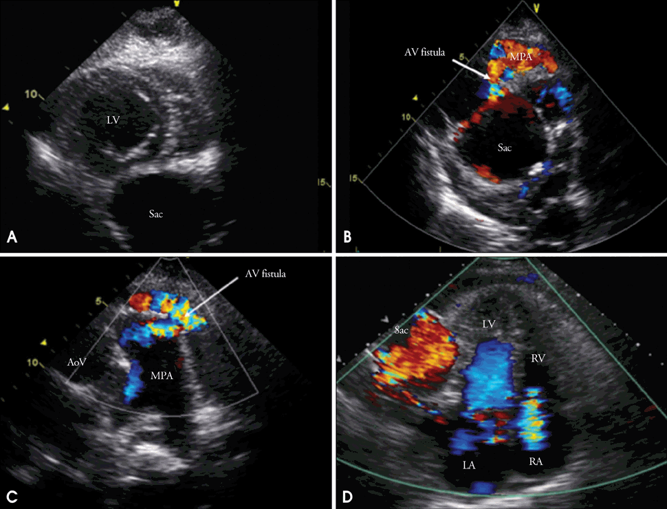
A: Echocardiogram in parasternal short axis view show extrinsic cardiac compression in left ventricle side due to multiple fusiform aneurysms (maximal diameter of largest one: about 6.7 cm). B: Modified apical 2 chamber view showed a fistula from aneurismal sac to main pulmonary artery. C: Parasternal short axis window in aortic valve level revealed a arteriovenous fistula from coronary artery to main pulmonary trunk. D: Apical 4 chamber view showed mild regurgitation of tricuspid valve. Sac: aneurysmal sac, LV: left ventricle, AoV: aortic valve, MPA: main pulmonary artery, AV: arteriovenous, LA: left atrium, RV: right ventricle, RA: right atrium.

Fig. 2.
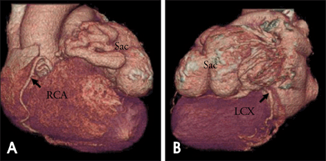
A 64-detector row cardiac computed tomography with 3D reconstruction showed several small tortous anonymous vessels from proximal right coronary artery (A) and enlarged tortous anonymous vessel from left main coronary artery along course of left circulflex artery (B). Arrow indicates RCA and LCX. Sac: aneurysmal sac, RCA: right coronary artery, LCX: left circumflex artery.

Fig. 3.
Coronary angiographic finding revealed left main coronary was dilated and communicated to aneurismal sac (A and B). And left anterior descending artery (LAD) was not visible. Small tortous anonymous vessel from proximal right coronary artery was drained to aneurysmal sac through a fistula formation (C).


 PDF
PDF ePub
ePub Citation
 Print
Print


 XML Download