

 PDF
PDF ePub
ePub Citation
 Print
Print


Introduction
Congenital pulmonary vein stenosis (CPVS) is one of cardiac anomalies producing generalized pulmonary venous hypertension such as mitral stenosis, a supravalvar mitral ring, mitral regurgitation, and total anomalous pulmonary venous connection.1) The prognosis is poor with the development of progressive pulmonary venous congestion followed by pulmonary arterial hypertension and eventual death.2)
The disorder is frequently overlooked during clinical examination and routine echocardiography because it is rare. Here, we report a patient with a history of cough, tachypnea and hemoptysis who was diagnosed with CPVS with a normal connection. The diagnosis was suspected based on an abnormal thoracic roentgenogram and confirmed by echocardiography, lung perfusion scanning, computerized tomography (CT) with angiogram, and cardiac catheterization with angiography.
Case
A 3 year old boy was admitted to Chonnam National University Hospital (CNUH) with a history of cough, tachypnea and hemoptysis. His parents visited a primary care physician due to the boy's persistent cough and exertional dyspnea. The pediatrician evaluated the plain radiographs, which revealed an enlarged pulmonary artery. The patient was transferred to CNUH with hemoptysis. On physical examination, his weight was 12 kg (3-10 percentiles) and height 94 cm (25-50 percentiles). His blood pressure was 90/40 mmHg (50 percentiles), pulse rate 106/min, respiratory rate 36/min and body temperature 36.2℃. On auscultation of the chest, decreased breathing sound of right lung field and an accentuated P2 without murmur were heard. Laboratory studies revealed hemoglobin of 10.5 g/dL, WBC of 8,600/mm3, (polymorphonuclear cell 22%, lymphocyte 65%, and monocyte 10%), platelet of 214,000/mm3, and oxygen saturation of 98%.
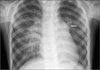
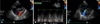
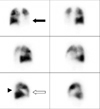
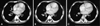
The thoracic roentgenogram showed dilated right pulmonary arteries and a pulmonary cornus with relative radiolucency at the left lower lung field, reticular interstitial infiltrations in the right mid portion and a normal sized heart (Fig. 1). The electrocardiogram revealed signs of right atrial enlargement and right ventricular hypertrophy with a sinus tachycardia. The two-dimensional and color Doppler echocardiography demonstrated pulmonary vein stenosis with a normally connected systemic venous connection. Three pulmonary veins appeared to be connected to the left atrium and continuously disturbed the pulmonary venous flow pattern; the peak velocity was 2.3 m/sec without phasic variation. The fourth pulmonary vein was not visible. The right atrium was enlarged and caused deviation of the interatrial septum toward the left atrium. The right ventricular pressure was increased with right ventricular enlargement. There were no associated cardiac malformations. There was moderate tricuspid regurgitation and a dilated main pulmonary artery consistent with severe pulmonary hypertension (Fig. 2). The Technetium-99m macro-aggregate lung perfusion scan showed absence of perfusion of the left lower lobe of the lung and diminished perfusion to the right middle and lower lobes of the lung (Fig. 3). The chest CT with angiogram showed interlobular septal thickening, which suggested interstitial pulmonary edema of the right middle lobe. The main pulmonary trunk and the right pulmonary artery were enlarged and the left pulmonary artery and its branches were observed to be small. The pulmonary vein that drained from the right middle lobe had focal stenosis. The one from the left upper lobe that drained the left atrium had relatively normal flow, but the pulmonary vein from the left lower lobe was not visible (Fig. 4).
The patient was diagnosed with a bilateral CPVS with a normal connection and treated with furosemide and aldactone to reduce the preload pressure on the heart. On about the 20th day after the first visit to CNUH, a cardiac catheterization was performed at Seoul National University Hospital according to the family's request. The cardiac catheterization showed a pulmonary artery pressure of 100/56 mmHg. The pulmonary artery wedge angiography revealed that the three pulmonary veins were stenosed (Fig. 5).
Discussion
Congenital pulmonary vein stenosis (CPVS) is a rare cardiac malformation especially with an anatomically normal connection.3) CPVS may occur as a focal stenosis of one or more veins at the atrial junction or a generalized hypoplasia characterized by narrowing of the lumen of the pulmonary veins for a considerable distance.1) Most commonly, both pulmonary veins from one lung are affected. However, Bini and colleagues reported patients with all of the pulmonary veins stenosed.3) The embryological basis for the stenosis of individual pulmonary veins appears to be abnormal incorporation of the common pulmonary vein into the left atrium.4) This developmental abnormality results in congenital annular fibrotic stenosis of the pulmonary venous orifice and extended constriction of the individual veins.4)
Stenosis of an isolated single pulmonary vein may not produce any symptoms. However, when several veins are involved, stenosis can cause significant clinical manifestations.1) The patients with CPVS and a normal connection are typically diagnosed as older infants and during childhood generally beyond the newborn period.3)5) They present with a long history of persistent tachypnea, dyspnea, recurrent pneumonia, and failure to thrive as well as cyanosis and hemoptysis.4)6-8) The symptoms ultimately progress to pulmonary edema and rightsided heart failure.3)4) However, Holcomb and associates reported a case of CPVS presenting as persistent pulmonary hypertension in the newborn.9)
The physical examination is consistent with the signs of pulmonary hypertension including a right ventricular lift, accentuation of the pulmonary component of the second heart sound and no significant murmurs. A pulmonary ejection click may be heard occasionally.1)4) CPVS is generally suspected by the chest radiographic findings. Asymmetric vascularity of both lung fields may be found, with increased vascularity of the unaffected lung segments.4)10) Additional findings include Kerley B lines, fluid in the fissures and interstitial edema of the affected lobes of the lungs.11) The heart is usually normal in size or slightly enlarged with right ventricular hypertrophy.1)4) The heart can shift toward the involved side with a prominence of the pulmonary trunk.1)4)11) Right ventricular hypertrophy and right atrial enlargement, without left-sided changes, is invariably found on the electrocardiography.1)4)
The Technetium-99m lung perfusion scan shows absence or diminished perfusion of the affected lung segments.4)11) Ventilation can be also be reduced in the same segments with variable degree; Kingston reported reduced ventilation in one patient with CPVS.12) Stenosis of the pulmonary veins can be recognized by two-dimensional echocardiography, magnetic resonance imaging or angiography. Discrete areas of narrowing or areas of hypoplasia may be noted in the distal pulmonary vein and at the pulmonary vein-left atrium junction, which are best imaged from the suprasternal, high parasternal or subcostal windows.4) The reflective capacity of the air in the lungs limits the ability of the ultrasound to provide images of the more proximal portions of the pulmonary veins.4) It may not be possible to demonstrate all four veins by precordial examination on the standard echocardiography, it is not a particularly better screening technique than transoesophagel color flow mapping or Doppler velocimetry.1)13) A diagnosis by standard echocardiography and Doppler methods is made more difficult by the distance of the pulmonary venous connection to the left atrium as measured by the transducer of the surface echocardiographic acoustic windows.13) However, the pulmonary veins are in close proximity to the transducer with transesophageal echocardiography.13) Pulsed Doppler echocardiography can be a useful tool for the evaluation of patients with suspected CPVS.9)14) Normal pulmonary venous flow is laminar and triphasic with the first and highest inflow during ventricular systole, the second inflow during the rapid filling phase of ventricular diastole and flow reversal after atrial contraction.9) However, stenotic pulmonary veins have a pattern of continuous flow that is disturbed without the normal phasic variation as well as turbulent flow.1)4)9)
Although abnormal pulmonary venous inflow patterns during Doppler echocardiography may suggest the presence of CPVS, a definitive diagnosis requires a cardiac catheterization.9) A pulmonary catheterization can be accomplished by the right side venous or left side arterial approaches.15) Although a trans-septal puncture is needed to enter the pulmonary veins, in the case of isolated pulmonary vein stenosis without interatrial communication, Bahl VK et al. describe an alternative retrograde non-trans-septal arterial approach for the pulmonary vein using steerable left-arterial catheter.15) The diagnostic features include the difference between the left atrial pressure and the pulmonary artery wedge pressure15) and the preferential flow occurring in the contralateral lung when unilateral pulmonary venous stenosis is associated with a left to right shunt.1)4) Pulmonary angiography shows the constriction of the affected pulmonary vein and the slow clearance of contrast medium from one lung in the case of unilateral pulmonary venous stenosis.1)4) Pulmonary arterial wedge angiography may be a better technique to demonstrate the precise anatomy of the pulmonary venous stenosis than pulmonary arteriography,1)16) where selective pulmonary venous injection is preferred especially in cases with isolated pulmonary vein stenosis.3)17) Both MRI and MRA are ideally suited for evaluating the pulmonary veins for stenosis4) even though Doppler echocardiography and cardiac catheterization have been regarded as the definitive diagnostic methods.9)
There is no effective treatment for severe pulmonary venous hypoplasia except pneumonectomy when the disease is unilateral. The treatment objective is to resolve the massive hemoptysis.1) The first surgical repair of a CPVS was reported by Kawashima and colleagues in 1971.18) Localized stenosis at the atrial junction has been treated in a few instances by patch grafting;19) however, diffuse restenosis has been documented as a significant cause of late mortality after repair.3) Due to the poor results of surgery for stenosis of the individual pulmonary veins, attempts to perform balloon angioplasty17) or cutting balloon angioplasty2) and utilization of balloon-dilatable stents have been reported.20)21) Results of both have been disappointing but the latter remains to be evaluated because of the possibility of reducing restenosis. The sutureless pericardial marsupialization procedure was introduced to minimize injury to the pulmonary vein wall by avoiding direct suturing of the veins, which appears superior to conventional techniques.22) Some physicians recommended lung or heart-lung transplantation for the rapidly progressive and uniformly fatal CPVS.5) Although long-term outcome from lung transplantation at a young age remains uncertain, all of the patients that underwent lung transplantation were alive and well at 6 to 24 months after transplantation.5)
The current case illustrates the rare malformation, a bilateral CPVS with normal connection. The two-dimensional and color Doppler echocardiography demonstrated the typical findings of CPVS in three veins. Cardiac catheterization ultimately revealed three stenosed pulmonary veins and pulmonary hypertension.
 XML Download