

 PDF
PDF ePub
ePub Citation
 Print
Print


Abstract
The purpose of this study was to evaluate the predictive value of ultrasonography (US) and 99mtechnetium dimercaptosuccinic acid (DMSA) scan for vesicoureteral reflux (VUR) in children with urinary tract infection (UTI). This study was a retrospective review of 114 children who were diagnosed as UTI from January 2004 to December 2007. A total of 114 patients underwent a US, DMSA scan and VCUG. The patients were divided into three groups according to the results of VCUG. The findings of the US and DMSA scan were compared with VCUG results. Of the 114 patients, there were 79 (69.3%) without VUR, 12 (10.5%) with low-grade VUR (grade I, II) and 23 (20.2%) with high-grade VUR (more than grade III). The US predicted 15 of 35 VUR with a sensitivity of 42.9% and a specificity of 70.9%. A DMSA scan predicted 26 of 35 VUR with a sensitivity of 74.3%. If either the US or DMSA scan was abnormal, this condition predicted 29 of 35 VUR with a sensitivity of 82.9%, negative predictive value (NPV) of 82.9%. VUR was associated with abnormal DMSA scan findings and abnormal findings of either US or DMSA scan. If either the US or DMSA renal scan was abnormal in children with a urinary tract infection, this was predictable factor for VUR. And this condition was more accurate in high-grade VUR. As screening examination for VUR, US and DMSA scan are useful and should be performed together. If both tests are normal in children with a urinary tract infection, there may be little or no clinically significant VUR.
Figures and Tables
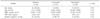
Table 3
Associations of each ultrasonographic finding with vesicoureteral reflux and high-grade vesicoureteral reflux
![]()
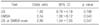
Table 4
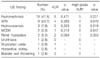
Associations of each 99mtechnetium dimercaptosuccinic acid scan finding with vesicoureteral reflux and high-grade vesicoureteral reflux
![]()
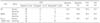
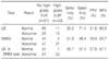
Table 6
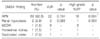
Relationship between abnormalities on image studies and the presence of vesicoureteral reflux
![]()
References
1. Elder Jack S. Kliegman RM, Behrman RE, Jenson HB, Stanton BF, editors. Urinary tract infection. Vesicoureteral reflux. Nelson textbook of pediatrics. 2007. 18th ed. Philadelphia: Saunders;2223–2233.
2. Bellinger MF, Duckett JW. Vesicoureteral reflux: a comparison of non-surgical and surgical management. Contrib Nephrol. 1984. 39:81–93.
3. Weiss R, Tamminen-Möbius T, Koskimies O, Olbing H, Smellie JM, Hirche H, et al. Characteristics at entry of children with severe primary vesicoureteral reflux recruited for a multicenter, international therapeutic trial comparing medical and surgical management. The International Reflux Study in Children. J Urol. 1992. 148:1644–1649.

4. Smellie JM, Ransley PG, Normand IC, Prescod N, Edwards D. Development of new renal scars: a collaborative study. Br Med J (Clin Res Ed). 1985. 290:1957–1960.
5. Jacobson SH, Hansson S, Jakobsson B. Vesico-ureteric reflux: occurrence and long-term risks. Acta Paediatr. 1999. 88:22–30.
6. Jakobsson B, Berg U, Svensson L. Renal scarring after acute pyelonephritis. Arch Dis Child. 1994. 70:111–115.
7. Wennerström M, Hansson S, Jodal U, Sixt R, Stokland E. Renal function 16 to 26 years after the first urinary tract infection in childhood. Arch Pediatr Adolesc Med. 2000. 154:339–345.
8. Smellie JM, Prescod NP, Shaw PJ, Risdon RA, Bryant TN. Childhood reflux and urinary tract infection: a follow-up of 10-41 years in 226 adults. Pediatr Nephrol. 1998. 12:727–736.
9. Jacobson SH, Eklöf O, Eriksson CG, Lins LE, Tidgren B, Winberg J. Development of hypertension and uraemia after pyelonephritis in childhood: 27 year follow up. BMJ. 1989. 299:703–706.
10. American academy of pediatrics. Committee on quality improvement. Subcomittee on urinary tract infection. Practice parameter: the diagnosis, treatment and evaluation of initial urinary tract infection in febrlie infants and young children. Pediatrics. 1999. 103:843–852.
11. Lavocat MP, Granjon D, Allard D, Gay C, Freycon MT, Dubois F. Imaging of pyelonephritis. Pediatr Radiol. 1997. 27:159–165.
12. Stashinko EE, Goldberger J. Test or trauma? The voiding cystourethrogram experience of young children. Issues Compr Pediatr Nurs. 1998. 21:85–96.
13. McAlister WH, Cacciarelli A, Shackelford GD. Complications associated with cystography in children. Radiology. 1974. 111:167–172.
14. Tseng MH, Lin WJ, Lo WT, Wang SR, Chu ML, Wang CC. Does a normal DMSA obviate the performance of voiding cystourethrography in evaluation of young children after their first urinary tract infection? J Pediatr. 2007. 150:96–99.
15. Oostenbrink R, van der Heijden AJ, Moons KG, Moll HA. Prediction of vesico-ureteric reflux in childhood urinary tract infection: a multivariate approach. Acta Paediatr. 2000. 89:806–810.
16. Lee SH, Noh SH, Oh JE, Kim MS, Lee DY. Predictive value for vesicoureteral reflux in children with urinary tract infection. J Korean Soc Pediatr Nephrol. 2008. 12:62–69.
17. Preda I, Jodal U, Sixt R, Stokland E, Hansson S. Normal dimercaptosuccinic acid scintigraphy makes voiding cystourethrography unnecessary after urinary tract infection. J Pediatr. 2007. 151:581–584.
18. Han SB, Ko YM, Lee SY, Jeong DC, Kang JH, Lee KY, et al. The significance of (99m)technetium dimercaptosuccinic acid(DMSA) scan as a substitute for voiding cystourethrography(VCUG) in evaluating children with first febrile urinary tract infection. J Korean Soc Pediatr Nephrol. 2007. 11:220–228.
19. Lee MD, Lin CC, Huang FY, Tsai TC, Huang CT, Tsai JD. Screening young children with a first febrile urinary tract infection for high-gradevesicoureteral reflux with renal ultrasound scanning and technetium-99m-labeled dimercaptosuccinic acid scanning. J Pediatr. 2009. 154:797–802.
20. Swerkersson S, Andreasson AC, Jodal U, Sixt R, Stokland E, Hansson S. The insignificance of low-grade vesicoureteral reflux [abstract]. Pediatr Nephrol. 2006. 10:1511A.
21. Goonasekera CD, Shah V, Wade AM, Barratt TM, Dillon MJ. 15-year follow-up of renin and blood pressure in reflux nephropathy. Lancet. 1996. 347:640–643.
22. Smellie JM, Barratt TM, Chantler C, Gordon I, Prescod NP, Ransley PG, et al. Medical versus surgical treatment in children with severe bilateral vesicoureteric reflux and bilateral nephropathy: a randomised trial. Lancet. 2001. 357:1329–1333.
23. Bailey RR, Lynn KL, Smith AH. Long-term followup of infants with gross vesicoureteral reflux. J Urol. 1992. 148:1709–1711.
24. Taskinen S, Rönnholm K. Post-pyelonephritic renal scars are not associated with vesicoureteral reflux in children. J Urol. 2005. 173:1345–1348.
25. Gordon I, Barkovics M, Pindoria S, Cole TJ, Woolf AS. Primary vesicoureteric reflux as a predictor of renal damage in children hospitalized with urinary tract infection: a systematic review and meta-analysis. J Am Soc Nephrol. 2003. 14:739–744.
26. Polito C, Rambaldi PF, Signoriello G, Mansi L, La Manna A. Permanent renal parenchymal defects after febrile UTI are closely associated with vesicoureteric reflux. Pediatr Nephrol. 2006. 21:521–526.
27. Ilyas M, Mastin ST, Richard GA. Age-related radiological imaging in children with acute pyelonephritis. Pediatr Nephrol. 2002. 17:30–34.
28. Jung SW, Jung KH, Kim MH, Lee JE, Hong YJ, Son BK. Factors associated with renal scarring in children with a first episode of febrile urinary tract infection. J Korean Soc Pediatr Nephrol. 2005. 9:56–63.
29. Goldman M, Bistritzer T, Horne T, Zoareft I, Aladjem M. The etiology of renal scars in infants with pyelonephritis and vesicoureteral reflux. Pediatr Nephrol. 2000. 14:385–388.
30. Polito C, Rambaldi PF, Mansi L, Di Toro R, La Manna A. Unilateral vesicoureteric reflux: low prevalence of contralateral renal damage. J Pediatr. 2001. 138:875–879.
31. Gleeson FV, Gordon I. Imaging in urinary tract infection. Arch Dis Child. 1991. 66:1282–1283.
32. Moorthy I, Easty M, McHugh K, Ridout D, Biassoni L, Gordon I. The presence of vesicoureteric reflux does not identify a population at risk for renal scarring following a first urinary tract infection. Arch Dis Child. 2005. 90:733–736.
33. Goldman M, Lahat E, Strauss S, Reisler G, Livne A, Gordin L, et al. Imaging after urinary tract infection in male neonates. Pediatrics. 2000. 105:1232–1235.
34. Schlesinger AE, Hernandez RJ, Zerin JM, Marks TI, Kelsch RC. Intero-bserver and intraobserver variations in sonographic renal length measure-ments in children. AJR Am J Roentgenol. 1991. 156:1029–1032.
35. Sargent MA, Long G, Karmali M, Cheng SM. Interobservervariation in the sonographic estimation of renal volume in children. Pediatr Radiol. 1997. 27:663–666.
36. Patel K, Charron M, Hoberman A, Brown ML, Rogers KD. Intra- and interobserver variability in interpretation of DMSA scans using a set of standardized criteria. Pediatr Radiol. 1993. 23:506–509.
37. Tsai JD, Huang FY, Tsai TC. Asymptomatic vesicoureteral reflux detected by neonatal ultrasonographic screening. Pediatr Nephrol. 1998. 12:206–209.
 XML Download