Abstract
MMP-1, EGF, and IL-1B gene polymorphism can be associated with gastric carcinogenesis. However, no study has yet confirmed the definitive role of these gene polymorphisms in gastric cancer risk. The 194 gastric cancer patients, 94 gastric adenoma patients, and 182 controls were used in this study. The SNP of the MMP-1 promoter, EGF, and IL-1B-31 were analyzed by PCR-RFLP and sequencing. The genotype frequency was compared between cases and controls, and a univariate and multivariate analysis were performed to determine the significant risk factors associated with gastric adenoma and adenocarcinoma. The frequency of 1G/2G genotypes in the MMP-1 promoter was similar to those in controls (p=0.734). The frequency of A/G genotypes in the EGF was similar to those in controls (p=0.239). The frequency of C/T genotypes in the IL-1B-31 was similar to those in controls (p=0.239). According to univariate analysis, male sex (p<0.0001), old age (≥60, p<0.0001), atrophy (pepsinogen I/II ≤3, p<0.0001), and G/G genotype of EGF (p=0.034) were significant risk factors associated with gastric adenoma and carcinoma. However, male sex (p=0.002), old age (p<0.0001), and atrophy (p<0.0001) were the only significant risk factors associated with gastric adenoma and carcinoma according to the multivariate analysis. In conclusion, the SNP of the MMP-1 promoter, EGF, and IL-1B-31 did not correlate with the risk of gastric adenoma and adenocarcinoma. Sex, age, and atrophy were the significant risk factors of gastric cancer.
Gastric cancer, the forth most common cancer and the second leading cause of cancer death in the world, has high incidence and mortality, particularly in Japan and Korea.1,2 Gastric carcinogenesis is a multistep process in which genetic and environmental factors interact with each other.3-7 Environmental factors, such as dietary habits, smoking, and helicobacter pylori infection are associated with higher risks of gastric cancer.7-9 Alterations in various genes, including oncogenes, tumor-suppressor genes, DNA repair genes, cell-cycle-related genes and cell-adhesion-related genes, have been implicated in the course of gastric carcinogenesis.10-12
Epidermal growth factor (EGF) activates multiple signaling pathways by binding with its receptor (EGFR),13,14 resulting in the proliferation, differentiation and tumorigenesis of epithelial tissues.15,16 Recently, a research suggested that A-G polymorphism of EGF might be involved, not only in the occurrence, but also in the malignant progression of gastric cancer.17 Matrix metalloproteinase-1 (MMP-1) plays a key role in cancer invasion and metastasis through the degradation of extracellular matrix (ECM) and basement membrane barriers.18 Helicobacter pylori encoding the pathogenicity island activates MMP-1 in gastric epithelial cells via JNK and ERK.19 The presence of 2G allele in the MMP-1 promotor has been reported to associate with the development and progression of colorecum cancer.20 Hamajima et al21 showed that the interleuking-1 (IL-1) gene polymorphism was associated with H. pylori persistent infection, which suggests that host genetic factors play a key role in susceptibility to H. pylori persistent infection. Among IL-1 Gene polymorphism, IL-1B-31T homozygotes have been related with the risk of intestinal types of gastric adenocarcinoma in the Korean population.22 MMP-1, EGF, and IL-1B gene polymorphism can be associated with gastric carcinogenesis. However, no study has yet confirmed the definitive role of these gene polymorphisms in gastric cancer risk.
In the present study, we investigated the genetic polymorphisms of MMP-1, EGF, and IL-1B with the risk of gastric adenoma and adenocarcinoma.
From March 2007 to December 2008, we recruited subjects (control group) who wished to receive a routine health checkup, including upper endoscopy, and a common screening examination for gastric cancer in Korea. All participants were interviewed for their medical and family histories. Subjects with a family history of gastric cancer, previous gastric adenoma and cancer were excluded. Patients who visited the gastroenterology and the surgery and endoscopy section of Chonnam National University Hospital, Gwangju, Korea were registered under control group, gastric adenoma (adenoma group) and adenocarcinoma (GC group). Before the examination, the purpose of the study was explained to the participants, and informed consent was obtained from all individuals. The institutional review board of Chonnam National University approved the protocol (CRI07081-3).
Peripheral blood drawn from a forearm vein was stored. Genomic DNA was isolated from whole blood from 194 of GC patients, 182 of control, 94 of adenoma patients by the alkaline lysis method using the QIAamp DNA Mini Kit (Qiagen inc. Valencia, CA, U.S.A).
1) MMP-1: The PCR primers used for amplifying MMP-1 polymorphism were: forward primer 5'-TGA CTT TTA AAA CAT AGT CTA TGT TCA-3'; reverse primer: 5'-TCT TGG ATT GAT TTG AGA TAA GTC ATA GC-3'. The reverse primer was specially designed to introduce a recognition site of restriction enzyme AluI (AGCT) by replacing a T with a G at the second position close to the 3' end of the primer. The 1G alleles have this recognition site, whereas the 2G alleles destroy the recognition site by inserting a guanine. Amplification of MMP-1 polymorphism was performed in a 25 µl reaction volume containing 200 ng of genomic DNA, and Tris-HCl (PH 9.0), (NH4)2SO4, 20 mM MgCl2, PCR enhancers in reaction buffer 2.5 µl, DW 16.5 µl, dNTP 0.5 µl, 10 pmol F and R primer 0.5 µl, DMSO (non specific band inhibitor) 2 µl, 1 unit Exprime Taq polymerase (Genet BIO Teageon, Korea) 0.5 µl.
DNA was predenatured at 94℃ for 10 min, and subjected to 35 cycles of denatured at 94℃ for 30 sec, and annealing at 58℃ for 30 sec, and extension at 72℃ for 30 sec followed by an extension of 72℃ for 10 min, and end at 4℃ for the final duration. Amplification products were resolved by electrophoresis on 1.8 % SeaKem LE agarose (Cambrex Bio Science Rockland Inc. Rockland, ME USA) next to a DNA MW standard marker 100 bp ladder (TAKARA BIO INC. Otsu, Shiga, Japan) and visualised with ethidium bromide staining. The MMP-1 amplification product size was 269 bp. The 269 bp fragment was the digested with Alu1 (TAKARA BIO INC. Otsu, Shiga, Japan) overnight at 37℃. After overnight digestion expected product sizes for alleles 2G, 1G, and 1G/2G are (269 bp), (241 bp and 28 bp), and (269 bp, 241 bp, and 28 bp) respectively.
2) EGF: Genotyping of EGF was done by PCR-RFLP as described previously. The PCR primers used for amplifying EGF polymorphism were: forward primer 5'-TGT CAC TAA AGG AAA GGA GGT-3'; reverse primer: 5'-TTC ACA GAG TTT AAC AGC CC-3'. Amplification of EGF polymorphism was performed in a 25 µl reaction volume containing 200 ng of genomic DNA, and Tris-HCl (PH 9.0), (NH4)2SO4, 20 mM MgCl2, PCR enhancers in reaction buffer 2.5 µl, DW 16.5 µl, dNTP 0.5 µl, 10 pmol F and R primer 0.5 µl, DMSO (non specific band inhibitor) 2 µl, 1 unit Exprime Taq polymerase (Genet BIO Teageon, Korea) 0.5 µl.
DNA was predenatured 94℃ for 10 min, and subjected to 35 cycles of denatured at 94℃ for 30 sec, and annealing at 58℃ for 30 sec, and extension at 72℃ for 1 min followed by an extension of 72℃ for 10 min, and end at 4℃ for the final duration. Amplification products were resolved by electrophoresis on 1.8% SeaKem LE agarose (Cambrex Bio Science Rockland Inc. Rockland, ME USA) next to a DNA MW standard marker 100 bp ladder (TAKARA BIO INC. Otsu, Shiga, Japan) and visualised with ethidium bromide staining. The EGF amplification product size was 242 bp. The 242 bp fragment was the digested with Alu1 (TAKARA BIO INC. Otsu, Shiga, Japan) overnight at 37℃. After overnight digestion expected product sizes for alleles G/G, A/A, and A/G are (193 bp, 34 bp, and 15 bp), (102 bp, 91 bp, 34 bp, and 15 bp), and (193 bp, 102 bp, 91 bp, 34 bp, and 15 bp) respectively.
3) IL-1B-31: The PCR primers used for amplifying IL-1B-31 polymorphism were: forward primer 5'-AGA AGC TTC CAC CAA TAC TC-3'; reverse primer: 5'-AGC ACC TAG TTG TAA GGA AG-3'. Amplification of IL-1B-31 polymorphism was performed in a 25 µl reaction volume containing 200ng of genomic DNA, and Tris-HCl (PH 9.0), (NH4)2SO4, 20 mM MgCl2, PCR enhancers in reaction buffer 2.5 µl, DW 16.5 µl, dNTP 0.5 µl, 10 pmol F and R primer 0.5 µl, DMSO (non specific band inhibitor) 2 µl, 1 unit Exprime Taq polymerase (Genet BIO Teageon, Korea) 0.5 µl.
DNA was predenatured at 94℃ for 10 min, and subjected to 35 cycles of denatured at 94℃ for 30 sec, and annealing at 55℃ for 30 sec, and extension at 72℃ for 1 min followed by an extension of 72℃ for 10min, and end at 4℃ for the final duration. Amplification products were resolved by electrophoresis on 1.8% SeaKem LE agarose (Cambrex Bio Science Rockland Inc. Rockland, ME USA) next to a DNA MW standard marker 100 bp ladder (TAKARA BIO INC. Otsu, Shiga, Japan) and visualised with ethidium bromide staining. The IL-1B-31 amplification product size was 239 bp. The 239 bp fragment was the digested with Alu1 (TAKARA BIO INC. Otsu, Shiga, Japan) overnight at 37℃. After overnight digestion expected product sizes for alleles C/C, T/T, and C/T are (239 bp), (137 bp and 102 bp), and (239 bp, 137 bp and 102 bp) respectively.
Serum anti-H. pylori antibody was measured using a commericial ELISA kit (Standard Diagnostics BIOLINE. Kyonggi-do, Korea). Seropositivity for H. pylori antibody was defined by optical density values according to manufacturer's protocol.
Serum pepsinogen was measured using high-performance liquid chromatography. Serum pepsinogen status was defined as atrophic when the criteria of both serum pepsinogen I level ≤70 ng/ml and a pepsignogen I/II ratio (serum pepsinogen I (ng/ml)/serum pepsinogen II (ng/ml)) ≤3.0 were simultaneously fulfilled.23
All statistical analysis was performed using statistical software package (SPSS 17.0 version for Windows, SPSS, Chicago, IL). Quantitative data were summarized as mean (standard deviation). One way ANOVA was used to compare the mean values of continuous variables, and a Kruskal-Wallis test was utilized for the comparison of discrete variables. A Univariate and multivariate logistic regression analysis were performed to assess the potential risk factors associated with gastric adenoma and carcinoma. A p value of less than 0.05 was accepted as statistically significant.
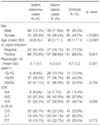
Baseline clinical characteristics of study subjects are summarized in Table 1. Among the GC cases, 71.6% were male versus 53.3% of the control (p<0.0001). The mean age (±SD) was 60.2 yr (±11.3) for GC cases and 46.1 yr (±11.0) for the control (p<0.0001). The man pepsinogen I/II ratio (±SD) was 4.3 (±3.4) for GC cases and 6.7 (±3.2) for the control (p=0.001). The frequency of 1G/2G genotypes in the MMP-1 promoter was similar to those in controls (p=0.734). The frequency of A/G genotypes in the EGF was similar to those in controls (p=0.239). The frequency of C/T genotypes in the IL-1B-31 was similar to those in controls (p=0.173). The genotype frequency of EGF in control group showed a significant deviation from Hardy-Weinberg equilibrium (p<0.05), but the others showed no significant deviation.
According to the univariate analysis, male sex (p<0.0001), old age (≥60, p<0.0001), atrophy (pepsinogen I/II ≤3, p<0.0001), and G/G genotype of EGF (p=0.034) were significant risk factors associated with gastric adenoma and carcinoma. However, H. pylori infection, IL-1B-31 Genotype, MMP-1 Genotype were not significant (Table 2). According to multivariate analysis, male sex (p=0.002), old age (p<0.0001), and atrophy (p<0.0001) were only significant risk factors associated with gastric adenoma and carcinoma (Table 3).
We assessed the association between the genetic polymorphisms of MMP-1, EGF, and IL-1B and the risk of developing gastric adenoma and adenocarcinoma, and found no significant association with 1G/2G genotypes in the MMP-1 promoter, A/G genotypes in the EGF, and C/T genotypes in the IL-1B-31.
Gastric carcinogenesis is a complex multifactorial and multistage process in which several factors are involved.24 Subjects infected with H. pylori are at an increased risk for developing gastric cancer, and cytokine gene polymorphisms represent one component of this complex process that may lead to the development of gastric cancer.24 The 1G/2G single nucleotide polymorphism (SNP) in the MMP-1 promoter at position -1,607 bp has been reported to affect the transcriptional activity. In the present study, the allelic frequency in the patients with gastric carcinoma was similar to that in controls, and the allelic frequency in the patients with gastric adenoma was also similar to that in controls. Therefore, it seems that the presence of 2G allele did not increase the susceptibility for the development of gastric carcinoma, especially in the early stage. This discrepancy between our results and previous results will be due to small sample number and ethnic differences.
EGF has many biological functions and plays an important role in the progression of various tumors, including gastric cancer. An A-G SNP at position 61 in the 5'-untranslated region (UTR) of the EGF gene has recently been reported to be associated with different levels of EGF production.17 In the present study, the alleleic frequency of A/G genotypes in the EGF with gastric adenoma and carcinoma was similar to that in controls. According to the univariate analysis, the G/G genotype of EGF was a significant risk factor associated with gastric adenoma and carcinoma, but the significance was absent in the multivariate analysis. Therefore, it seems that the presence of 2G allele of EGF did not increase the susceptibility for the development of gastric carcinoma, especially in the early stages. This discrepancy between our results and previous results will be due to small sample number and ethnic differences.
In the current study, there were no significant differences in the allelic frequency of IL-1B-31 among gastric adenoma, carcinoma and control. Recent meta-analysis showed that only the IL1RN×22 Genotype seems to consistently increase the risk of gastric precancerous lesions, supporting a role for this polymorphism in the early stages of gastric carcinogenesis.25 Therefore, it seems that the presence of C/T genotypes in the IL-1B-31 did not increase the susceptibility for the development of gastric carcinoma, especially in early stages. This discrepancy between our results and previous results will be due to small sample number and ethnic differences.
In the present study, male sex (p=0.002), old age (p<0.0001), and atrophy (p<0.0001) were the only significant risk factors associated with gastric adenoma and carcinoma according to the multivariate analysis. These results indicate that the patients with these factors can be at a high risk group for the development of gastric cancer. High risk groups merit intensive gastric cancer screening.
In conclusion, the SNP of the MMP-1 promoter, EGF, and IL-1B-31 did not enhance the risk of gastric cancer. Sex, age, and atrophy were the only significant risk factors in the development of gastric cancer.
Figures and Tables
Acknowledgement
This work was supported by a research grant from the Research Institute of Medical Sciences, Chonnam National University (2007-CURIMS-DR009).
References
4. Correa P. Human gastric carcinogenesis: a multistep and multipfactorial process -First American Cancer Society Award Lecture on Cancer Epidemiology and Prevention. Cancer Res. 1992. 52:6735–6740.
5. Tahara E. Molecular mechanism of stomach carcinogenesis. J Cancer Res Clin Oncol. 1993. 119:265–272.

6. Stadtlander CT, Waterbor JW. Molecular epidemiology, pathogenesis and prevention of gastric cancer. Carcinogenesis. 1999. 20:2195–2208.

7. Neugut AI, Hayek M, Howe G. Epidemiology of gastric cancer. Semin Oncol. 1996. 23:281–291.
8. Kelly JR, Duggan JM. Gastric cancer epidemiology and risk factors. J Clin Epidemiol. 2003. 56:1–9.

9. Malaty HM, Engstrand L, Pedersen NL, Graham DY. Helicobacter pylori infection: genetic and environmental influences. A study of twins. Ann Intern Med. 1994. 120:982–986.

10. Yasui W, Yokozaki H, Fujimoto J, Naka K, Kuniyasu H, Tahara E. Genetic and epigenetic alterations in multistep carcinogenesis of the stomach. J Gastroenterol. 2000. 35:111–115.
11. Werner M, Becker KF, Keller G, Höfler H. Gastric adenocarcinoma: pathomorphology and molecular pathology. J Cancer Res Clin Oncol. 2001. 127:207–216.

12. González CA, Sala N, Capellá G. Genetic susceptibility and gastric cancer risk. Int J Cancer. 2002. 100:249–260.

13. Jorissen RN, Walker F, Pouliot N, Garrett TP, Ward CW, Burgess AW. Epidermal growth factor receptor: mechanisms of activation and signaling. Exp Cell Res. 2003. 284:31–53.

14. Olayioye MA, Neve RM, Lane HA, Hynes NE. The ErbB signaling network: receptor heterodimerization in development and cancer. EMBO J. 2000. 19:3159–3167.

15. Laurence DJ, Gusterson BA. The epidermal growth factor. A review of structural and functional relationships in the normal organism and in cancer cells. Tumour Biol. 1990. 11:229–261.
16. Singletary SE, Baker FL, Spitzer G, Tucker SL, Tomasovic B, Brock WA, et al. Biological effect of epidermal growth factor on the in vitro growth of human tumors. Cancer Res. 1987. 47:403–406.
17. Hamai Y, Matsumura S, Matsusaki K, Kitadai Y, Yoshida K, Yamaguchi Y, et al. A single nucleotide polymorphism in the 5' untranslated region of the EGF gene is associated with occurrence and malignant progression of gastric cancer. Pathobiology. 2005. 72:133–138.

18. Kohn EC, Liotta LA. Molecular insights into cancer invasion: strategies for prevention and intervention. Cancer Res. 1995. 55:1856–1862.
19. Krueger S, Hundertmark T, Kalinski T, Peitz U, Wex T, Malfertheiner P, et al. Helicobacter pylori encoding the pathogenicity island activates matrix metalloproteinase 1 in gastric epithelial cells via JNK and ERK. J Biol Chem. 2006. 281:2868–2875.

20. Ghilardi G, Biondi ML, Mangoni J, Leviti S, DeMonti M, Guarnelline E, et al. Matrix metalloproteinase-1 promotor polymorphism 1G/2G is correlated with colorectal cancer invasiveness. Clin Cancer Res. 2001. 7:2344–2346.
21. Hamajima N, Matuso K, Saito T, Tajima K, Okuma K, Yamao K, et al. Interleukin 1 polymorphisms, lifestyle factors, and Helicobacter pylori infection. Jpn J Cancer Res. 2001. 92:383–389.

22. Chang YW, Jang JY, Kim NH, Lee JW, Lee HJ, Jung WW, et al. Interleukin-1B (IL-1B) polymorphisms and gastric mucosal levels of IL-1Beta cytokine in Korean patients with gastric cancer. Int J Cancer. 2005. 114:465–471.

23. Watabe H, Mitsushima T, Yamaji Y, Okamoto M, Wata R, Kokubo T, et al. Predicting the development of gastric cancer from combining Helicobacter pylori antibodies and serum pepsinogen status: a prospective endoscopic cohort study. GUT. 2005. 54:764–768.


 PDF
PDF ePub
ePub Citation
 Print
Print


 XML Download