

 PDF
PDF ePub
ePub Citation
 Print
Print


Introduction
Surgical correction of lower eyelid retraction frequently requires a spacer graft between the tarsus and the lower eyelid retractor. Available spacers include donor sclera, ear cartilage, autogenous hard palate, polyester mesh, and porous polyethylene.1-5 Silicone sponges have long been used as implants in scleral buckling, and they have appropriate hardness and elasticity for use as spacers.6 We describe a case of lower eyelid retraction after inferior rectus (IR) disinsertion in a patient with congenital fibrosis of the extraocular muscles (CFEOM), which was corrected with silicone sponge spacers.
Case Report
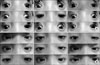
A 5-year-old boy presented with bilateral fixed downgaze with exotropia and chin elevation. The patient had a history of bilateral ptosis and limitation of eye movements after birth. On examination, his corrected visual acuity was 10/20 in both eyes and he had 25 prism diopters (PD) of hypotropia and 30 PD of exotropia bilaterally in the primary position, with chin elevation. The patient could abduct his eyes slightly, but he was unable to upgaze, downgaze, or adduct (Fig. 1A), and he had no levator function. The patient was diagnosed clinically with CFEOM. We performed disinsertion of the lateral rectus (LR) and IR in both eyes and did not secure the LR and IR to the sclera. We also recessed the inferior and lateral conjunctivae by 5 mm. After the operation, the patient's chin elevation improved, and his eye position was orthophoria in the primary position. However, the patient showed eyelid retraction by 3 mm from the inferior corneal limbus in the right eye and 2 mm in the left eye, along with inferior conjunctival hyperemia (Fig. 1B).
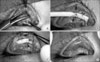
To correct the lower eyelid retraction, we horizontally dissected the inferior conjunctival fornix with a monopolar coagulator (Ellman Surgitron, Ellman International Inc., Hewlett, NY, USA) and separated the lower eyelid retractor from the lower tarsal plate. After further dissection of the orbital fat pad, we inserted a silicone sponge (MIRA #504; diameter - 4 mm, length - 15 mm) between the inferior tarsal plate and the lower eyelid retractor and sutured it to the inferior border of the tarsal plate and the lower eyelid retractor with 6-0 Vicryl (Fig. 2).
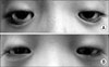
At the 3-month postoperative visit, both lower eyelid margins were at the inferior corneal limbus in the primary position, and there was no restriction of lower eyelid movement. No complications were noted 1 year after the operation, and eyelid retraction did not recur (Fig. 3).
Discussion
CFEOM is characterized by congenital nonprogressive bilateral ptosis and restrictive external ophthalmoplegia, with the eyes usually fixed in a hypotropic position.7-9 CFEOM has historically been considered a primary myopathy, in which there is fibrosis of the ocular musculature and fibrotic adhesions between Tenon's capsule and the extraocular muscles.7 According to recent studies, the myopathic changes are secondary to a primary innervation defect or aberrant innervation of the extraocular muscles caused by genetic mutations.8,9 CFEOM is characterized by congenital bilateral ptosis and ocular motility dysfunction, with the globes infraducted in the primary position and with restricted upgaze and variably restricted horizontal gaze. Our patient had these characteristics and had fibrotic adhesions between Tenon's capsule and the extraocular muscles that were noted intraoperatively.
Management of CFEOM consists of amblyopia treatment, correction of vertical and horizontal misalignment, and correction of blepharoptosis. To correct vertical misalignme nt, IR recession can be performed with or without superior rectus resection. Because our patient had globe restriction after IR and LR detachment, we did not secure the IR and the LR to the sclera. When large IR recession or disinsertion is performed, lower eyelid retraction occurs through the action of the check ligaments between the IR and the tarsus of the lower eyelid, such as with our patient.
Lower eyelid retraction results in cosmetic problems, ocular discomfort, and exposure keratitis. Surgical management frequently requires a spacer graft between the inferior tarsal plate and the lower eyelid retractor. To date, several materials have been used as spacers, including autogenous hard palate mucosa, donor sclera, and ear cartilage, as well as synthetic materials like polyester mesh, porous polyethylene, and polytetrafluoroethylene.1-5,10 These materials have both advantages and significant disadvantages. Autogenous hard palate mucosa is associated with donor site morbidity, and its preparation is time-consuming. Homologous grafts such as donor sclera carry a risk of disease transmission and graft absorption. Autogenous ear cartilage grafts cause relative immobility of the lower eyelid. Polyester mesh is not rigid enough for support, and porous polyethylene is associated with implant exposure, unexplained pain, and poor eyelid mobility on downgaze.2,5 Polytetrafluoroethylene has many advantages; it is easily cut, molded, and sutured and is resistant to infection, but the pore size is too small for rapid ingrowth of fibrovascular tissue, and it carries the risk of exposure.6 Ideal spacers must have appropriate thickness and rigidity and must resist rejection and absorption.
In this case, we used a silicone sponge for a spacer; this has not been previously reported for the correction of lower lid retraction. Silicone sponges have long been used as implants for scleral buckling, but they also have the appropriate hardness and elasticity required for spacers.10 Silicone is highly biocompatible and incites little inflammatory response. Silicone sponges have the appropriate hardness for lower eyelid support and the proper elasticity for enabling free lower eyelid movement. They also have adequately sized pores for ingrowth of fibrovascular tissue and are easily cut, molded, and sutured. Preoperative soaking in antibiotics can decrease the risk of postoperative infection, and meticulous trimming and suturing to the conjunctiva and Tenon's capsule can prevent silicone sponge exposure. In our case, there were no complications or recurrence 1 year after the operation.
This case illustrates the successful use of silicone sponges as spacers for the correction of lower lid retraction in CFEOM after strabismus surgery. Silicone sponges might also be considered for the lid retraction associated with Graves' ophthalmopathy, facial palsy, trauma, and cicatrization.
 XML Download