

 PDF
PDF ePub
ePub Citation
 Print
Print


Introduction
A coronary artery fistula is an anomaly between a coronary artery and one of the cardiac chambers, great vessels, or another vascular structure.1,2 Coronary artery fistulas are not common but are clinically important in adulthood because of an increased risk of complications, such as heart failure, myocardial ischemia, arrhythmias, infective endocarditis, and rupture.1 We present a case report of a patient with two huge coronary arteriovenous fistulas with aneurysmal changes diagnosed by echocardiography and angiography and detailed by 64-slice multidetector computed tomography (MDCT).
Case Report
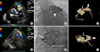
A 72-year-old woman presented with a short history of exertional dyspnea. Her medical history included type 2 diabetes mellitus (DM), hypertension, atrial fibrillation, and moderate mitral regurgitation (MR). She had been complaining of chronic fatigue and dizziness of unidentified origin for years. Her other past history included a subtotal thyroidectomy for thyroid cancer. Chest radiography showed marked cardiomegaly with a large amount of left pleural effusion. An electrocardiogram revealed atrial fibrillation with a rapid ventricular response. On physical examination, her blood pressure was 114/53 mm Hg and chest auscultation revealed continuous murmurs over the left upper sternal border and apex. A 2D-echocardiogram revealed bilaterally enlarged atria, moderate MR, and a dilated right upper pulmonary vein drainage site. Accentuation of abnormal color Doppler flows around the pulmonary trunk and the left ventricle were observed (Fig. 1 A, B). Coronary angiography showed two huge coronary arteriovenous fistulas with aneurysmal changes, one of which originated from the dilated main artery of the left pulmonary trunk (Fig. 1 C) and another from the right coronary artery adjacent to the pulmonary trunk (Fig. 1 D). We subsequently performed 64-slice multi-detector CT angiography to determine the anatomy in detail, which demonstrated a markedly dilated left main artery with an arteriovenous fistula from the left main artery of the pulmonary trunk and an abnormal vessel originating from the right coronary cusp adjacent to the origin of the right coronary artery drained to the dilated, aneurysmal left main artery (Fig. 1 E, F). The patient underwent surgical ligation of both coronary arteriovenous fistulas after receiving therapy for symptoms and signs of congestive heart failure. Her dyspnea resolved with surgery.
Discussion
Coronary artery fistulas are rare.1 Angiographic series present an incidence of 0.3~0.8%.1 The reported incidence of coronary-pulmonary artery fistulas constitutes 15~20% of all coronary artery fistulas, and they are associated with aneurysmal dilatation in 19~26% of cases.3 Most of these fistulas arise from the right coronary artery or from the left anterior descending artery. The circumflex coronary artery is rarely involved.1
Most adult patients are asymptomatic, but some patients present with exertional dyspnea, chest pain, syncope, and palpitation, presumably due to shunt-induced reduction in coronary blood flow.1,3,4 With large fistulas, half of the patients develop complications, such as congestive heart failure, infective endocarditis, rupture of an aneurysm, or myocardial infarction.5
The traditional diagnostic tool for coronary artery fistulas has been coronary angiography, but considerable progress has been made in the field of noninvasive coronary imaging.2 New imaging modalities such as MDCT have been used to diagnose coronary artery fistulas with more accurate detail of the anatomy of the abnormal coronary artery, which facilitates the best management strategies, as described in this case.2-4,6
Coronary computed tomography angiography (CTA) is a relatively new imaging modality that has been used for coronary artery imaging since 2000.6 Previous systems had limited spatial and temporal resolution and were compromised by image-noise.6 However, with the introduction MDCT, the image quality was improved.6 Coronary CTA demonstrates accurate views of the cardiac chambers, coronary arteries, and coronary veins.6 Because of its excellent spatial resolution and ability to demonstrate relationships between anatomical structures, coronary CTA may be the appropriate diagnostic modality for coronary artery fistulas, especially for fistulas coursing between the coronary artery and other vascular structures, and to detect fistula origin and drainage sites.2-4,6 Coronary CTA would be an ideal noninvasive technique for imaging in patients with suspected coronary artery fistulas.4,6
 XML Download