

 PDF
PDF ePub
ePub Citation
 Print
Print


INTRODUCTION
The Clonal expansion of donor T cells, which respond to the recipient's environment, causes acute graft-versus-host disease (aGVHD) and graft-versus-leukemic (GVL) effects during early immune reconstitution after transplantation. The pathophysiology of aGVHD is thought to be due to donor T cell recognition of anti-genic differences by antigen-presenting cells (APCs), activated cytokine storm and mediated cellular and inflammatory responses on the target tissue.1,2 Several animal studies have suggested that down-regulation of aGVHD and establishment of immune tolerance are mediated by antigen-specific regulatory T cells (Tregs).3-7
Tregs play a key role in hematopoietic stem cell transplantation (HSCT); they prevent aGVHD and promote engraftment and chimerism by specific induction of tolerance of the alloantigens. The expression of FOXP3, which encodes a forkhead/winged helix transcription factor and is required for the development and the functional activity of CD4+CD25+ Tregs, is used as a reliable molecular marker for quantifying Tregs in the peripheral blood.8-10 CD4+CD25+ Tregs can block the amplification of the NK cell response to APCs, and depletion of Tregs can result in the enhancement of NK cell-mediated anti-tumor immunity.11,12 Importantly, several in vitro studies have reported that the suppression of proliferation and function of cytotoxic T cells by Tregs results in an unfavorable influence on allogeneic transplantation interfering with the GVL activity of donor T cells.13-15 However, conflicting results have been reported where inhibition of GVHD by Tregs did not interfere with the GVL effects after allogeneic HSCT.16,17
In this study, we evaluated whether the expression of CD4+CD25+ Tregs and NK cells is associated with the incidence of aGVHD or whether Tregs have a differentiated potential between GVL effects and aGVHD without causing disease relapse during early immune reconstitution after myeloablative HSCT.
Materials and Methods
1. Patients and transplant approach
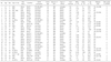
This study was conducted between September 2002 and January 2007 after obtaining informed consent and the approval from our institutional review board. Twenty-nine patients who underwent non-T cell-depleted myeloablative allogeneic HSCT were analyzed to investigate the relationship between early reconstitution of Tregs, NK cells, the incidence of aGVHD, and disease relapse. The clinical characteristics of the patients are shown in Table 1.
All patients received non-T cell-depleted grafts after myeloablative conditioning with fludarabine (180 mg/m2) and busulfan (12.8 mg/kg) (n=13), cyclophosphamide (120 mg/kg) and total body irradiation (1,200 cGy) (n=7), or busulfan (16 mg/kg) and cyclophosphamide (120 mg/kg) (n=9). Nine patients received stem cells from unrelated donors and five patients were infused from HLA-mismatched donors. Three of five patients with HLA-mismatched HSCT had alemtuzumab (30 mg) added to the conditioning to reduce aGVHD. Cyclosporin A (CSA) or FK-506 was used for prophylaxis against GVHD, starting on day -3, continued for 180 days, and then tapered over a 90-day period. A short course of methotrexate was added to the protocol in patients without alemtuzumab conditioning.
2. Cell isolation
Peripheral blood samples were obtained at 21 days after HSCT until the absolute neutrophil count reached above 1,000×109/L. Peripheral blood mononuclear cells (PBMC) were separated from blood samples by Ficoll-Hypaque (lymphoprep; 1.077 density) gradient centrifugation and were frozen in RPMI 1,640 media with 20% fetal bovine serum and 10% dimethyl sulfoxide. Before analysis, frozen cells were thawed, washed and resuspended. The level of FOXP3 mRNA expression was assessed by quantitative real-time reverse transcription PCR.
3. Flow cytometry
Immunophenotyping was performed on collected peripheral blood samples. Briefly, mononuclear cells isolated by Ficoll-Hypaque density gradient centrifugation were incubated with the following mouse monoclonal antibodies: Rhodamine 1-conjugated anti-human CD16, CD56 and fluorescein isothiocyanate (FITC)-conjugated anti-CD3, CD4-phycoerythrin (PE), and CD25-FITC (Coulter Immnuotech, Miami, FL, USA). CD4+CD25+ T cells in PBMCs were sorted on a BD FACS Aria cell sorter (BD Biosciences, San Jose, CA 95131, USA) using PE-conjugated anti-CD4, FITC-conjugated anti-CD25, and its isotype control antibody. The level of FOXP3 mRNA expression was measured in sorted CD4+CD25+T cells. At least 5,000 cells were counted and analyzed by use of BD FACS Aria cell sorter software.
4. RNA extraction and real-time reverse transcription PCR for FOXP3
Total RNA from cells was isolated by using TRIzol (Invitrogen, Karlsruhe, Germany). The first-strand cDNA was synthesized from 100 ng of total RNA by use of a SuperScript III kit (Invitrogen) according to the manufacturer's protocol. Human FOXP3 mRNA expression was quantified by using a SYBR green quantitative PCR kit (Takara, Japan) with the Rotor-gene 3,000 (CORBETT, Australia) and was corrected by amplification of a control, the human β-actin housekeeping gene. Amplification was carried out in a total volume of 20 µL for 40 cycles of 15 s at 95℃, 20 s at 60℃ and 20 s at 72℃. The samples were run in triplicate, and their relative expression was determined by normalizing the expression of each target gene to β-actin and then comparing this normalized value to the normalized expression in a reference sample from which the fold change was calculated. For FOXP3, the following primer combinations were used: forward, 5'-CGG ACA CTC AAT GAG ATC TA-3'; and reverse, 5'-ATC CTC CTT TCC TTG ATC TT-3'. The FOXP3 primers were synthesized by Integrated DNA Technologies. For β-actin, the primers were as follows: forward, 5'-GAT GAG ATT GGC ATG GCT TT-3'; reverse, 5'-CAC CTT CAC CGT TCC AGT TT -3'.
5. Statistical Analysis
To compare the level of FOXP3 expressions and the expression of NK cells between grade 0-1 aGVHD and grade 2-4 aGVHD or between relapse and no relapse, discrete or continuous variables were analyzed by use of the Fisher's exact t test and Mann-Whitney U test, respectively. p values <0.05 were considered statistically significant.
Results
We studied all 29 patients who underwent non-T cell-depleted myeloablative allogeneic HSCT and analyzed the correlation between Tregs and immunophenotypic expression of NK cells at early engraftment. In addition, we analyzed whether the level of FOXP3 expressions differed in relapsed cases compared with cases who did not relapse. The population of Tregs in CD4+ CD25+ T cells was evaluated by real-time quantitative PCR for FOXP3 gene expression.
The median age of the patients was 34 years (range, 16~53 years) and there were more males than females. The median follow-up time was 17.2 months (range, 6.6~57.8 months). Fourteen patients developed grade 2~4 aGVHD and 13 patients experienced relapse. The median dose of infused CD34+ cells was 4.18×10⁶/kg, and T cell doses were 34.4×10⁷/kg. Only one patient, who had leukemic marrow before transplantation, failed to achieve an engraftment. Unrelated donor, HLA mismatching and alemtuzumab conditioning were significantly related to the incidence of aGVHD (p<0.05). Alemtuzumab conditioning and aGVHD were strongly associated with relapse. Patients with grade 2~4 aGVHD had a significantly lower relapse rate (n=1, 7.1%) than did those with grade 0~1 aGVHD (n=12, 80%) (p=0.00). The overall median level of FOXP3 expression and the percentage of NK cells for all patients was a median 6.38 ng/µL (range, 0.36~19.87 ng/µL) and 19.8% (range, 2.0~60.0%), respectively.
1. The level of FOXP3 expression in CD4+CD25+ T cells correlates with aGVHD and a relapse in early engraftment
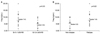
The expressions of the FOXP3 gene in CD4+CD25+ T cells showed that patients with grade 2~4 aGVHD (median, 5.36 ng/µL; range, 0.36~10.31 ng/µL) had significantly lower levels of FOXP3 gene expression than did those with grade 0~1 aGVHD (median, 7.45 ng/µL; range, 1.40~19.87 ng/µL; p=0.03) (Fig. 1A). However, the level of FOXP3 gene expressions in patients with relapse (median, 7.46 ng/µL; range, 3.42~19.87 ng/µL) was significantly higher than in those without relapse (median, 5.52 ng/µL; range 0.36~10.31 ng/µL) (p=0.02) (Fig. 1B). The three patients treated with alemtuzumab conditioning had no aGVHD; their level of FOXP3 expression was a median of 8.91 ng/µL (range, 4.76~12.31 ng/µL). They all relapsed after a median of 6.6 months (range, 1.1~31.4 months) from transplantation (p=0.04).
2. No inverse relationship between Tregs and NK cells and no correlation between NK cells and aGVHD or a relapse
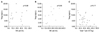
When we investigated populations of NK cells during early engraftment, we found that the percentage of NK cells in patients with grade 2~4 aGVHD was a median of 22.2% (range, 2.0~60.0%) and that in patients with grade 0~1 aGVHD was a median of 18.3% (range, 2.6~42.6%; p=0.55). There was no significant difference in the population of NK cells in the relapsed (median, 28.3%; range, 2.6~60.0%) and non-relapsed patients (median, 18.0%; range, 2.0~48.1%) nor between the two aGVHD groups (p=0.51). The analysis of the influence of Tregs on the activation of NK cells, during the early post-transplantation period, showed that NK cell proliferation in response to FOXP3 expression, with or without aGVHD, was not influenced by early reconstitution of CD4+FOXP3+ regulatory T cells (Fig. 2A, B). In addition, the grafted donor T-cell doses did not affect the level of FOXP3 gene expression during the early post-HSCT period (p=0.18) (Fig. 2C).
Discussion
After hematopoietic stem cell transplantation, the key donor immune cells controlling early transplant outcomes (eg, GVHD and relapse) are the transplanted nonthymic-dependent donor T cells and early recovering NK cells because thymic function is defective or absent in adults, broad cell-mediated immune recovery occurs months to years after transplantation.18,19 The early post-transplant period is characterized by powerful immune reactions causing GVHD and GVL. To identify the GVHD-reacting T cells and to separate from those conferring GVL, in vitro and in vivo studies have suggested that distinct subsets of host-reacting and leukemia-reacting T cells separate GVHD from GVL alloreactivity.20-22 This selective biology of GVHD and GVL activity has been investigated including the evaluation of regulatory T cells in allogeneic HSCT. Recent studies by Trenado et al16 and Edinger et al17 suggested that CD4+CD25+ regulatory T cells might preserve the GVL activity while suppressing GVHD in a mouse model of transplantation. However, conflicting studies have reported that regulatory T cells may reduce anti-tumor immunity in murine models and in human subjects.23-25
In the present study, the early reconstitution of Tregs after HSCT was significantly related with the development of aGVHD. This finding is consistent with recent published studies.26,27 However, we did not confirm a role for Tregs in the suppression of the proliferation of NK cells or differential effects on aGVHD or GVL effects. This might be because of the different rate of relapse between the grade 0~1 aGVHD group (80%) and the grade 2~4 aGVHD group (7.1%). In addition, the three patients who were treated with the alemtuzumab conditioning had relatively high levels of FOXP3 expression and experienced relapse without aGVHD. Alemtuzumab is used to deplete recipient lymphocytes in vivo, thereby enhancing engraftment, and to deplete donor T cells to reduce the incidence of aGVHD. The affect of alemtuzumab on immunosuppression after HSCT is estimated to persist for over three months.28 However, alemtuzumab conditioning did not suppress the reconstitution of CD4+CD25+ Tregs during early engraftment and did not cause grade 2~4 aGVHD in this study, although a limited number of patients were evaluated. We demonstrated that alemtuzumab conditioning was associated with CD4+CD25+ Tregs after early HSCT and the severity of aGVHD as well as the risk of relapse.
Several studies29-31 reported on how donor T cells affect engraftment after HSCT and prevent the rejection of stem cells. Regulatory T-cell content, in the grafted donor T cells, may be part of an important mechanism underlying engraftment by inducing donor tolerance and preventing aGVHD. The demonstration that Tregs could separate aGVHD from GVL activity suggested that their immunosuppressive potential could be manipulated to reduce aGVHD without detrimental consequences on GVL effects. However, it remains unclear whether the early-engrafted Tregs have the same features at the time of relapse. Hence, just before relapse, patients have both donor-type and recipient-type Tregs in mixed chimerism. No studies have determined which type of Tregs play a dominant role in causing a relapse.
In summary, our findings suggest that CD4+CD25+ Tregs during early engraftment have an inverse correlation with the risk of aGVHD and a direct correlation with relapse. However, we failed to identify a correlation between the expression of CD4+CD25+ Tregs and NK cells in patients with non-T cell-depleted allogeneic HSCT.
 XML Download