

 PDF
PDF ePub
ePub Citation
 Print
Print


Introduction
Heart failure (HF) is a clinical syndrome that occurs in patients who, because of an inherited or acquired abnormality of cardiac structure or function, develop a constellation of clinical symptoms (dyspnea and fatigue) and signs (edema and rales) that lead to frequent hospitalization, a poor quality of life, and a shortened life expectancy.1 HF constitutes one of the leading worldwide causes of morbidity and mortality. More than 20 million people are affected with HF. The general prevalence of HF in the adult population in developed countries is about 2%. The prevalence is continuously growing, however, especially in developed countries. Because current therapies for cardiac disorders, such as acute myocardial infarction (AMI), valvular heart disease, and arrhythmias, are allowing patients to survive longer, almost all forms of cardiac diseases can be translated to HF. In developed countries, coronary artery disease (CAD) has become the major etiology in 60~75% of HF. In 20~30% of the cases of HF with a depressed ejection fraction (EF), the exact etiologic basis is not known. These patients are referred to as having non-ischemic, dilated, or idiopathic cardiomyopathy (CMP) if the cause is unknown. Despite many recent advances in the evaluation and management of HF, including anti-HF medications that have reasonable effects against HF, e.g., β-adrenergic receptor blockers, angiotensin-converting-enzyme inhibitor (ACEI), angiotensin receptor blocker (ARB), aldosterone blocking diuretics, mechanical ventricular assist devices, cardiac resynchronization, and ultimately cardiac transplantation, the development of symptomatic HF carries a poor prognosis. About 30~40% of patients die within 1 year of diagnosis and 60~70% die within 5 years, mainly from worsening HF or as a sudden event. Therefore, we need more specific treatment options to decrease the morbidity and mortality of HF.
In the recent 2~3 decades, gene therapy (GT) has been continuously exploited and recommended to control HF. Cardiovascular GT is a novel technology for treating cardiovascular diseases by replacing defective or missing gene products, including cellular proteins, receptors, and inhibitors. GT shifts the therapeutic focus toward correcting the pathophysiology at the cellular and subcellular levels. There are many technological obstacles to progress in gene therapy for HF. Such obstacles include the discovery of reasonable target molecules and genes in HF development and aggravation, the development of less toxic and immunogenic gene transferring systems, and effective and safe gene expression and protein manufacturing in the host. Gene therapies still have various fabulous benefits to apply to HF treatment. Currently, the candidate genetic targets of GT for HF are intracellular calcium regulation and homeostasis, β-adrenergic receptor signaling, antiapoptotic signaling, and myocardial angiogenesis.2-5 Generally, viral vector systems have been the major gene transferring system in this field, although microRNA and biocompatible polymer vector systems have also been introduced recently.6 General methods of GT using intracellular Ca2+ handling for heart failure are depicted.7 The purpose of this article is to supply some knowledge about GT as a potential treatment for HF. In this review, we attempt to explain the mechanism and usefulness of the GTs that are related to calcium handling in the cardiomyocytes (CMCs) in the HF.
Gene transferring system into cardiomyocytes
1. Intracoronary catheter delivery
Intracoronary catheter gene delivery is a relatively safe, easy to apply method in GT for HF. Generally used diagnostic or interventional coronary artery catheters and a programmable infusion pump system can be used for this procedure. Compared with thoracotomy and cardiopulmonary bypass, this method is simple and safe, even in patients with severely depressed cardiac function. But, this method is generally inefficient for myocardial gene transfer unless certain adjuvants and specific conditions are used. In an early phase clinical trial using an adeno-associated virus (AAV) 1 vector containing a single-strand 4486 nucleotide DNA expressing the human sarco/endoplasmic reticulum Ca2+ATPase2a (SERCA2a), an intracoronary catheter-based gene delivery method was used.8
2. Intraventricular delivery
The most global and efficient gene delivery method in a preclinical, small animal HF model is the intraventricular injection via the apical myocardium to the aortic root with aortic and pulmonary artery clamping for 10~40 seconds.9 Gene spreading to the entire myocardium with this method is very efficient compared with other delivery methods. This method could be applied to human subjects with HF in a limited manner under the cardiopulmonary bypass applied for other conditions, e.g., coronary bypass or open heart surgery.
3. Retroperfusion via the coronary sinus
Catheterization into a coronary sinus or subselective cardiac vein under the concurrent occlusion of outflow from the coronary sinus distributes the transgene in a retrograde fashion from the coronary venous circulation through the capillary bed to the CMC and interstitium in potentially all regions of the myocardium.10 Compared with intramyocardial injection, this method results in more homogeneous transmural gene expression.10
4. Intramyocardial direct injection
Direct injection of the genetic material into a myocardium can be done under beating heart surgery or cardioplegia with cardiac bypass with thoracotomy. Local expression and invasiveness limit its use. Moreover, the needle track in the myocardium can be an arrhythmogenic focus. To overcome these limitations of myocardial gene injection, catheter-based endomyocardial gene injection using a nonfluoroscopic, three-dimensional mapping and injection (NOGA) system has been applied in the clinical field. This method was also adopted in the study of Adenovirus (Adv)-Vascular Endothelial Growth Factor (VEGF)121 GT for non-optional CAD and some stem cell injection for HF.11 Even with the use of endomyocardial catheter-based gene delivery, however, local expression of genetic material is a big disadvantage of this method, especially in patients with global ventricular dysfunction.
5. Intrapericardial gene delivery
After the injection of genetic material into the pericardial space, 25% of it is recovered in the myocardium. To elevate myocardial transfer efficiency, a thermosensitive gel/plasmid DNA mixture can be used. This formulation improves retention time at the site of injection and enhances myocardial delivery efficiency with low cytotoxicity compared with a PEI/DNA mixture.12 This method can be applied to humans with a small thoracotomy or video-assist thoracostomy.
6. Percutaneous cardiac recirculation-mediated gene transfer
Recirculating perfusate delivery was achieved with the use of a novel cardiac perfusion circuit in this method.13 Under fluoroscopic guidance, coronary venous blood is recaptured from the coronary sinus with the use of a percutaneously positioned occlusive balloon recovery catheter. The draining catheter is placed in such a position to exclude the azygous vein, either by occluding it with the balloon or by incorporating a structural element beyond the catheter to prevent dynamic collapse during the application of suction. Venous return is facilitated by the use of a roller pump, followed by reoxygenation with an oxygenator membrane. The oxygenated perfusate is then directed to the left coronary territory via a nonocclusive catheter placed percutaneously in the left main coronary artery. To determine the distribution of the myocardial perfusate delivered in this manner, we characterized the pattern of delivery of the fluorophore indocyanine green (ICG) in 10 milliliters of circuit recirculated for 10 min. The heart was then explanted and examined by using near-infrared spectroscopy.
Promising gene therapy molecular targets for heart failure
1. Calcium handling pathways in the cardiomyocyte
Distorted handling of Ca2+ in the CMCs is a hallmark of HF. There is a decreased sarcoplasmic reticulum (SR) Ca2+ content and a prolonged Ca2+ transient, which is generally considered to be a consequence of increased Na+-Ca2+ exchanger (NCX). Also present are a reduction of SERCA2a, a decreased phospholamban (PLN)/SERCA2a ratio, and an augmented open probability of the ryanodine receptor (RyR).14 In addition to these changes causing dysfunctional contractile performance, the decreased clearance of the cytosol Ca2+ may increase the risk of arrhythmias and may also precipitate pathological cardiac remodeling.15
2. SR Ca2+-ATPase (SERCA2a)
SERCA2a is the cardiac isoform of this family of Ca2+-ATPase, and a loss of its activity and resultant decrease in SR Ca2+ uptake is a feature of the failing CMCs, including in human HF.16 SERCA2a activity in myocytes is tightly controlled by PLN, a small inhibitory peptide that regulates SERCA2a on a beat-to-beat basis. The inhibitory action of PLN is set by its phosphorylation status.17 Failing human CMCs showed recovery of impaired Ca2+ homeostasis and normalization of dysfunctional contractile responses after SERCA2a GT.17 Intracoronary Adv-SERCA2a delivery to rats in HF resulting from transaortic constriction (TAC) led to improved systolic and diastolic function along with dramatically improved survival 4 weeks after GT.18 GT with SERCA2a was approved for human heart failure in a phase I/II trial (CUPID trial).11 A total of 9 patients with advanced HF received a single intracoronary infusion of AAV1/SERCA2a in the open-label study. Doses administered ranged from 1.4×10¹¹ to 3×10¹² DNase resistant particles per patients. Patients were followed up for 6 to 12 months. Several patients demonstrated improvements from baseline to month 6 across a number of parameters of HF, including symptomatic, functional, biomarker, and LV functioning/remodeling. Two patients who failed to improve had preexisting anti-AAV1 neutralizing antibodies.19
3. Phospholamban (PLN)
PLN plays an important role in the regulation of SR Ca2+ homeostasis by mediating the break system for SERCA2a. Overexpression of PLN in rabbits provokes CMP.20 PLN-overexpressing transgenic mice show Ca2+ handling derangement and CMP.21 PLN gene knockout (KO) mice result in enhanced ventricular contractility and a blunted response to β-adrenergic stimulation.22 In rats with cardiac dysfunction due to MI, the AAV-antisense PLN gene delivery to the myocardium around the MI effectively attenuated the depression of cardiac dysfunction, significantly inhibited PLN expression, and enhanced myocardial SERCA acitivity.23 Tsuji et al. reported that antisense PLN rescued infarcted rat hearts from LV contractile dysfunction associated with an unchanged myocardial oxygen consumption (VO2) intercept of the VO2-systolic pressure volume area linear relation. Therefore, antisense PLN prevents Ca2+ overload-induced LV contractile dysfunction in terms of mechanical work and especially energetics.24 Adv-antisense PLN gene delivered to myocardial cells obtained from human end stage heart failure decreased PLN expression over 48 hours, increasing contraction and relaxation velocity. PLN restored the frequency response in the failing CMCs to normal, and increasing frequency resulted in enhanced SR Ca2+ release and contraction.25 Despite these beneficial effects of PLN ablation, a naturally occurring PLN KO mutation resulted in a severe LV contraction disorder.26 Genetically ablated PLN could have significant differences in biological effect compared with acute lowering of the PLN level in a HF model. More elegant studies are necessary to confirm this controversy.
4. Protein Phsosphatase (PP) 1 and Inhibitor Protein (IP)-1
The action of PLN on SERCA2a is subject to secondary control mediated by protein phosphatase (PP) 1 dephosphorylation. PP1 action is also controlled by the action of phosphatase inhibitors, inhibitor protein (IP) I-1 and I-2. A polymorphism in IP I-1 (G147D) is common in HF patients, exclusively in black subjects. Adv-D147 (mutant type) GT to human CMCs showed no significant differences in fractional shortening or contraction or relaxation rates, but blunted contractile responses after isoproterenol stimulation compared with Adv-G147 (wild type) infection.27 Adv-IP I-2 delivery to inhibit PP1 action in genetic CMP in hamsters improved LV fractional shortening and reduced LV chamber size at 1 week. This GT significantly extended the survival time of cardiomyopathic hamsters for 3 months.28 Transgenic mice with heart-directed overexpression of a constitutively active form of IP I-2 under the TAC for 28 days exhibited a higher atrial mass and an enhanced ventricular m-RNA expression of β-myosin heavy chain. This transgenic mouse showed reduced LV contractility and depressed shortening of isolated myocytes. This phenomenon was originated from a profound abnormal cytosolic Ca2+ transient and a reduced phosphorylation of PLN after TAC in transgenic mice.29 According to the above data, GT targeted to PP1 or IP expression, which induces SERCA2a activity boosting, could be good strategy in HF in the future.
5. Parvalbumin (PV)
PV is an acidic, intracellular Ca2+ binding protein of low molecular weight. It is associated with several Ca2+-mediated cellular activities and physiologic responses. PV may function as a "Ca2+ shuttle" that transports Ca2+ from troponin-C to the SR Ca2+ pump during muscle relaxation. PV supplied to CMCs offers an energy-independent removal of cytosolic available Ca2+ and correction of the prolonged diastolic Ca2+ decay generally seen in HF without further energy deprivation. PV gene transfer with Adv vector to Dahl salt-sensitive rats, which have diastolic dysfunction, caused significantly faster relaxation. This result was more evident with β-subtype PV than with α-subtype PV. This difference originated from the predominant affinity of the β-subtype PV to Ca2+ and Mg+ related to cytosolic Ca2+ traffic.30 Adv-PV GT to senescent and adult CMCs obtained from F344/BN hybrid rats increased the rate of myocyte re-lengthening and Ca2+ transient decay without an effect on myocyte shortening.31 This favorable effect of PV was confirmed in vivo. Adv-PV GT to aged F344/BN rats reduced LV diastolic pressure and the time course of pressure decline but had no effect on systolic parameters. This result shows that PV GT may address impaired Ca2+ homeostasis and diastolic dysfunction without an increase in energy expenditure.32 Selectivity of PV Ca2+ binding to the relaxation phase of the Ca2+ cycle is determined by the rate of Ca2+ influx and expression levels of PV. In the gene transfer of PV, there is some complex dose-response between the sarcomere length shortening-frequency and PV concentration in adult rat CMCs in primary culture. At low estimated PV (approximately 0.01 mM), contractile parameters were unchanged; at intermediate PC, the relaxation rate of the mechanical contraction and the decay rate of the Ca2+ transient increased with little effects on amplitude; and at high PV (approximately 0.1 mM), the relaxation rate was further increased, but the amplitudes of the mechanical contraction and the calcium transient were diminished compared with control myocytes. Therefore, there is an optimal PV range for which the relaxation rate is increased with little effect on contractile amplitude and PV effectiveness decreases as the stimulus frequency increases.33 Further investigation of physiologic responses along with PV expression and tissue level in an optimal HF animal model and human CMCs is essential to apply these potentially beneficial findings to human HF.
6. S100A1
S100A1, a Ca2+-binding protein of the EF-hand type, is preferentially expressed in the healthy heart and has been identified as a positive inotropic regulator of in vivo and in vitro myocardial contractility with cardioprotective actions at least in vitro. S100A1 undergoes a large conformational change upon binding Ca2+ as necessary to interact with numerous protein targets. Targets of S100A1 include proteins involved in Ca2+ signaling (RyR, SERCA2a, PLN) and Ca2+-activated proteins (annexin V/VI, S100B, S100A4, A100P, and F1 ATP synthase).34 S100A1 expression levels in myocardial tissue are significantly down-regulated in end-stage HF. Intracoronary delivery of the human S100A1 gene by the use of a first-generation Adv normalized the diminished S100A1 protein abundance in a rat HF model that in turn rescued both impaired contractile performance and Ca2+ cycling of the failing myocardium.35 Adv-S100A1 gene delivery into rat CMCs increased fractional shortening by 55% and systolic Ca2+ amplitudes by 62%, whereas S100A1 protein increased SERCA activity by 28%. The gain in systolic Ca2+ supply was not only seen during basal conditions but also with isoproterenol-stimulated Ca2+ cycling. Thus, S100A1 enhances cardiac contractility by increasing intracellular Ca2+ fluxes at least in part due to a modulation of SERCA.36 above result was confirmed with transgenic overexpression of the S100A1 in mouse. Contractile function and Ca2+ handling properties were increased in ventricular myocytes isolated from S100A1 transgenic mice. Enhanced cellular Ca2+ cycling by S100A1 was associated both with increased SR Ca2+ content and with enhanced SR Ca2+-induced Ca2+ release, and S100A1 was shown to be associated with the cardiac RyR.37 AAV6-S100A1 delivery with aortic root injection under ascending aortic clamping showed improved SR Ca2+ handling, including improved contractile function and LV remodeling in rat HF.38 The use of S100A1 as a therapeutic target is actually not restricted to cardiac GT but could be employed in the emerging fields of cardiac tissue engineering and cell-based therapy. Engineered heart tissue from rat CMCs showed enhanced contractile performance at baseline and in response to β-adrenergic receptor stimulation after S100A1 gene addition, indicating that S100A1-based genetic manipulation might be a promising strategy to strengthen the therapeutic effectiveness of artificial cardiac tissue or even artificial hearts.39 Further evidence gathering from large animal HF models with S100A1 GT is essential to apply this potential therapeutic molecule to human HF.
Gene transfer vectors
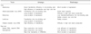
Both viral and non-viral vector systems are available. But, there is no exist one vector system meets all the various criteria, e.g., ease of preparing, excellent gene transfection efficiency, durable gene expression, and noninflammatory and nonimmunogenic properties. Recently, a non-viral vector system has been under investigation to improve gene transfection efficiency with low immunogenicity. The major vector systems are summarized in Table 1.
1. Adenoviral (Adv) vector
The Adv vector is the most commonly applied vector in preclinical HF models because of its high gene transfer efficiency into nondividing cells like CMCs. The disadvantages of the Adv vector are its relatively short duration of gene expression (3~4 weeks), neutralizing antibody induction (which is related to a reduction of gene expression in the case of repeated injection), immunogenicity, and precipitation of cellular apoptosis or necrosis.40 The new-generation Adv vectors shows longer gene expression and reduced immunogenicity.40
2. Adeno-associated virus (AAV) vector
AAV vectors effectively transfer genetic material to both dividing and nondividing cells and they integrate into the genome of the host cell and support durable transgenic expression for about 30 weeks.41 However, it is very difficult to generate a purified high viral titer with an AAV vector and its packaging capability is less than 4.7 kilobases.42
3. Retroviral and lentiviral vectors
Retroviral vectors are less immunogenic and their transfection efficiency is good in dividing cells in vitro. Long-term genetic expression is guaranteed, but transfection efficiency to nondividing cells is very low. The lentiviral vector may achieve efficient expression in nondividing cells or beating cells like CMCs.43
4. Nonviral vector systems
Nonviral vectors are relatively safe but inefficient for gene transfer to the myocardium. Examples of nonviral vectors include naked plasmid DNA, gene gun (using bioballistic bombardment of DNA-coated gold particles to penetrate target organs or single cell layer), plasmid-liposome complexes (lipoplex), and liposome-polymer-DNA complexes (lipoplex). Unlike viral vector DNA, internalization of naked DNA or DNA conjugates can occur either in a random manner or upon binding to a specific receptor depending on the design of conjugates.44 A controlled-release DNA-polymer coating delivery system has been investigated for transfection of cardiac tissue.45
Recently, synthetic nonviral materials are fast gaining popularity as safe and efficient vectors for delivering genes to target organs. Not only can nanoparticles function as efficient gene carriers, they also can simultaneously carry diagnostic probes for direct "real-time" visualization of gene transfer and downstream process.46 For example, ORMOSIL (organically modified silica) nanoparticles can condense, protect, and deliver plasmid DNA within cells. This nanoparticle was effectively used in mouse brain tissue with stereotactic surgery.47 Nonviral vector systems are fascinating in the aspect of low immunogenicity and no association with virus-related infection, but low gene transfer efficiency is still a major problem in the use of an in vivo system.
In summary, some hurdles remain to improving treatment efficacy for human HF. The molecular and cellular mechanisms of HF should be exploited continuously and vigorously. For most human HF cases, etiologies are very complex and multigenetic problems are anticipated. Elegant studies for enlightening diverse genetic abnormalities and molecular pathologies for initiation and aggravation of HF must be conducted. Also, the well-known etiologic diseases for HF, e.g., hypertension, diabetes mellitus, ischemic heart disease, and valvular heart disease, should be treated thoroughly. For treating heart failure, new drugs should be invented and manufactured. Otherwise, we must have continuous interest in new therapeutic options, e.g., gene therapy or stem cell therapy because of the incompleteness of classic HF treatment options. There are many obstacles to advancing GT for HF, e.g., searching for powerful candidate genes; preparing gene transfer vectors that have excellent packaging properties, transfection efficiency, low inflammation, and low immunogenicity; and developing appropriate small and large animal models of HF. Currently, a few of the targets discussed above are in preclinical stages with the goal of clinical trials in the near future. The initial safe and relatively acceptable outcomes of the human early stage clinical trial with AAV-SERCA2a (the CUPID study) are enormously valuable. This result will speed the human clinical trials of GT for HF.
 XML Download