

 PDF
PDF ePub
ePub Citation
 Print
Print


Abstract
Sphingosine-1-phosphate (S1P), a bioactive sphingolipid metaolite, regulates multiple cellular responses such as Ca2+ signaling, cellular growth and survival, and differentiation. Sphingosine kinase (SphK) is a key enzyme in modulating the levels of S1P and is emerging as an important regulating enzyme. Here we have investigated whether K6PC-5, a newly synthesized SphK activator, plays a neuroprotective role by activating cell survival systems such as protein kinase C, phosphatidylinositol-3-kinase (PI3K), and acting on the anti-apoptotic and the pro-apoptotic genes in SN4741 dopaminergic cells. 1-methyl-4-phenylpyridinium is a neurotoxin that selectively inhibits the mitochondrial functions of dopaminergic neurons in the substantia nigra pars compacta. In the present study, we found that MPP+ induced a decrease in SN4741 mouse dopaminergic cell viability. K6PC-5 restored the reduced phospho-PKC and phospho-PI3K activities caused by MPP+ toxicity. In addition, gene expression analysis revealed that K6PC-5 prevented both the MPP+- induced expression of the pro-apoptotic gene mRNA, Bax, and the decrease of the anti-apoptotic gene mRNA, bcl-w. These results suggest that the neuroprotective mechanism of K6PC-5 against MPP+-induced apoptotic cell death includes stimulation of PKC and PI3K, and modulation of cell survival and death genes.
Figures and Tables
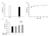 | Fig. 1MTT+ neurotoxicity and protection by K6PC-5 in SN4741 mouse dopaminergic neuronal cell line as measured by MTT reduction assay (A, B, and C). (A) Compare to media treated cells, treatment of cells with DMSO showed no significant decrease in the cell viability. (B) The viability of SN4741 cells was increased with increasing concentration of K6PC-5. (C) Protective effect of K6PC-5 against dopaminergic neuronal cell death in SN4741 cells. SN4741 cells were pretreated with varying concentrations of K6PC-5 (0.1, 1, 5 uM) for 1 hr, followed by incubation with 1 mM MPP+ for 24 hr. The viability was markedly reduced by a 24 hr treatment with 1 mM MPP+, whereas 1 hr pretreatment with K6PC-5 (0.1, 1, 5 uM) conferred significant protection against MPP+-induced neurotoxicity. These data represent the mean±SE (standard error) of three independent experiments, with each point done in quadruplicate.
|
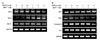 | Fig. 2Modulation of anti- and pro-apoptotic gene expressions by K6PC-5 and MPP+. SN4741 cells were treated with K6PC-5 (0.1, 1, 5 uM), MPP+ (1 mM), or the combination of the two compounds for 6 hr. Cellular RNA from each treatment was extracted and the mRNA expression for Bcl-2, Bcl-w, Bcl-xl, Bad, Bax, Caspase-6, p21, and GAPDH was analyzed by RT-PCR.
|
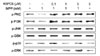 | Fig. 3Effects of K6PC-5 and MPP+ on p-PKC, p-PI3K, p-JNK, p-GSK, p-p38, and p-ERK immunoreactivities in SN4741 cells. Cells were treated with K6PC-5 (0.1, 1, 5 uM), MPP+ (1 mM), or the combination of the two drugs for 3 hr. Cell lysis buffer extracted cellular proteins from each treatment.Toxicity test of MPP+ (A) and EGCG (B) compounds in SN4741 mouse dopaminergic neuronal cell line. In vitro cytotoxic cell death for MPP+ and EGCG were assessed by MTT reduction assay in SN4741 cells at 24h.
|
References
1. Gerlach M, Ben-Shachar D, Riederer P, Youdim MB. Altered brain metabolism of iron as a cause of neurodegenerative diseases. J Neurochem. 1994. 63:793–807.

2. Jenner P, Olanow CW. Oxidative stress and the pathogenesis of Parkinson's disease. Neurology. 1996. 47:6 Suppl 3. S161–S170.
3. Jenner P. Oxidative stress in Parkinson's disease. Ann Neurol. 2003. 53:Suppl 3. S26–S38.
4. Dauer W, Przedborski S. Parkinson's disease: mechanisms and models. Neuron. 2003. 39:889–909.
5. Noh HG, Jang SJ, Park SJ, Kim SH, Jeong HS, Park JS. Protective effects of epigallocatechin-3-gallate against 1-methyl-4-phenylpyridinium-induced dopaminergic neuronal cell death. Chonnam Med J. 2007. 43:73–79.
6. Krishnamurthi R, Stott S, Maingay M, Faull RL, McCarthy D, Gluckman P, et al. N-terminal tripeptide of IGF-1 improves functional deficits after 6-OHDA lesion in rats. Neuroreport. 2004. 15:1601–1604.
7. Schober A. Classic toxin-induced animal models of Parkinson's disease: 6-OHDA and MPTP. Cell Tissue Res. 2004. 318:215–224.
8. Ryu EJ, Angelastro JM, Greene LA. Analysis of gene expression changes in a cellular model of Parkinson disease. Neurobiol Dis. 2005. 18:54–74.
9. Huwiler A, Kolter T, Pfeilschifter J, Sandhoff K. Physiology and pathophysiology of sphingolipid metabolism and signaling. Biochim Biophys Acta. 2000. 1485:63–99.
10. Zhang YH, Vasko MR, Nicol GD. Intracellular sphingosine 1-phosphate mediates the increased excitability produced by nerve growth factor in rat sensory neurons. J Physiol. 2006. 575(Pt 1):101–113.
11. Liu H, Chakravarty D, Maceyka M, Milstien S, Spiegel S. Sphingosine kinases: a novel family of lipid kinases. Prog Nucleic Acid Res Mol Biol. 2002. 71:493–511.
12. Shu X, Wu W, Mosteller RD, Broek D. Sphingosine kinase mediates vascular endothelial growth factor-induced activation of ras and mitogen-activated protein kinases. Mol Cell Biol. 2002. 22:7758–7768.
13. Edsall LC, Cuvillier O, Twitty S, Spiegel S, Milstien S. Sphingosine kinase expression regulates apoptosis and caspase activation in PC12 cells. J Neurochem. 2001. 76:1573–1584.
14. Taha TA, Hannun YA, Obeid LM. Sphingosine kinase: biochemical and cellular regulation and role in disease. J Biochem Mol Biol. 2006. 39:113–131.
15. Olivera A, Kohama T, Edsall L, Nava V, Cuvillier O, Poulton S, et al. Sphingosine kinase expression increases intracellular sphingosine-1-phosphate and promotes cell growth and survival. J Cell Biol. 1999. 147:545–558.
16. Spiegel S, Milstien S. Sphingosine-1-phosphate: an enigmatic signalling lipid. Nat Rev Mol Cell Biol. 2003. 4:397–407.
17. Youm JK, Jo H, Hong JH, Shin DM, Kwon MJ, Jeong SK, et al. K6PC-5, a sphingosine kinase activator, induces anti-aging effects in intrinsically aged skin through intracellular Ca(2+) signaling. J Dermatol Sci. 2008. 51:89–102.
18. Cuvillier O, Pirianov G, Kleuser B, Vanek PG, Coso OA, Gutkind S, et al. Suppression of ceramide-mediated programmed cell death by sphingosine-1-phosphate. Nature. 1996. 381:800–803.
19. Xia P, Wang L, Moretti PA, Albanese N, Chai F, Pitson SM, et al. Sphingosine kinase interacts with TRAF2 and dissects tumor necrosis factor-alpha signaling. J Biol Chem. 2002. 277:7996–8003.
20. Cordey M, Gundimeda U, Gopalakrishna R, Pike CJ. Estrogen activates protein kinase C in neurons: role in neuroprotection. J Neurochem. 2003. 84:1340–1348.
21. Levites Y, Amit T, Youdim MBH, Mandel S. Involvement of protein kinase C activation and cell survival/ cell cycle genes in green tea polyphenol (-)-epigallocatechin 3-gallate neuroprotective action. J Biol Chem. 2002. 277:30574–30580.
22. Gubina E, Rinaudo MS, Szallasi Z, Blumberg PM, Mufson RA. Overexpression of protein kinase C isoform epsilon but not delta in human interleukin-3-dependent cells suppresses apoptosis and induces bcl-2 expression. Blood. 1998. 91:823–829.
 XML Download