Abstract
To evaluate the therapeutic effects of 0.05% cylosporine A eyedrops (Restasis, Allergan Inc., Irvine, CA, USA.) in patients with dry eye syndrome, one hundred thirty-five patients (270 eyes) with dry eye syndrome were treated with 0.05% cylosporine A (CsA) and lubrication eyedrops for 12 months. Before and 1, 3, 6 and 12 months after treatment, symptom score, corneal sensitivity, tear film break-up time (BUT), keratoepitheliopathy score, basal tear secretion, tear clearance rate were measured. Before and 3 months after treatment, conjunctival impression cytology were done. One month after treatment, symptom score (2.38±0.80), basal tear secretion (6.34±3.26 mm), and keratoepitheliopathy score (1.10±1.54) were improved from 2.87±0.62 (p〈0.01), 5.89±2.72 mm (p〈0.01) and 1.50±1.82 (p〈0.01), respectively. Three months after treatment, tear film break-up time (5.70±1.76 sec) and tear clearance test (13.52±11.0) were improved from 5.24± 2.01 sec (p=0.01) and 10.74±8.48 (p=0.02), respectively. Six months after treatment, corneal sensitivity were improved from 57.85±4.27 mm to 57.57±6.24 mm (p=0.01) during the follow up period. Treatment of dry eye syndrome with topical CsA resulted in an increase in goblet cell numbers and decrease in conjunctival keratinization. Fourteen patients (10.4%) were discontinued instillation of topical CsA due to burning sensation and pain. Use of 0.05% topical CsA emulsion is effective for dry eye syndrome in helping to improve ocular symptom, and parameters of tear film and ocular surface during the follow-up period of 12 months.
Figures and Tables
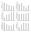
 | Fig. 1Tear film and ocular surface changes after treatment with topical 0.05% cyclosporine A in patients with severe dry eye. (A) symptom score, (B) corneal sensitivity, (C) basal secretion test, (D) tear film break-up time, (E) keratoepitheliopathy score, (F) tear clearance rate. *p <0.05 compared with baseline. |
 | Fig. 2Slit lamp photographs with fluorescein staining in dry eye syndrome patient before (A) and after (B)topical 0.05% cyclosporine A therapy. Note the marked improvement in the corneal epithelium. |
 | Fig. 3Impression cytologic finding (PAS, × 400). (A) Specimen from a patients with dry eye before treatment of 0.05% topical cyclosporin application shows a goblet cell loss and large, polygonal epithelial cells with a nucleocytoplasmic ratio of 1 : 3. (B) Specimen at three months after 0.05% topical cyclosporine application show many periodic acid-schiff positive goblet cells and round epithelial cells with a nucleocytoplasmic ratio of 1 : 1. |
References
1. Tang-Liu DD, Acheampong A. Ocular pharmacokinetics and safety of ciclosporin, a novel topical treatment for dry eye. Clin Pharmacokinet. 2005. 44:247–261.

2. Dick AD, Azim M, Forrester JV. Immunosuppressive therapy for chronic uveitis: optimising therapy with steroids and cyclosporin A. Br J Ophthalmol. 1997. 81:1107–1112.

3. Stevenson D, Tauber J, Reis BL. The Cyclosporin A Phase 2 Study Group. Efficacy and safety of cyclosporin A ophthalmic emulsion in the treatment of moderate-to-severe dry eye disease: a dose-ranging, randomized trial. Ophthalmology. 2000. 107:967–974.

4. Miyata K, Amano S, Sawa M, Nishida T. A novel grading method for superficial punctate keratopathy magnitude and its correlation with cornea epithelial permeability. Arch Ophthalmol. 2003. 121:1537–1539.

5. Anshu , Munshi MM, Sathe V, Ganar A. Conjunctival impression cytology in contact lens wearers. Cytopathology. 2001. 12:314–320.

7. Yoon KC, Heo H, Im SK, You IC, Kim YH, Park YG. Comparison of autologous serum and umbilical cord serum eyedrops for dry eye syndrome. Am J Ophthalmol. 2007. 144:86–92.
8. Dick AD, Azim M, Forrester JV. Immunosuppressive therapy for chronic uveitis: optimising therapy with steroids and cyclosporin A. Br J Ophthalmol. 1997. 81:1107–1112.

9. Tatlipinar S, Akpek EK. Topical ciclosporin in the treatment of ocular surface disorders. Br J Ophthalmol. 2005. 89:1363–1367.

10. Her J, Yu SI, Seo SG. Clinical effects of various antiinflammatory therapies in dry eye syndrome. J Korean Ophthalmol Soc. 2006. 47:1901–1910.
11. Kaan G, özden ö. Therapeutic use of topical cyclosporine. Ann Ophthalmol. 1993. 25:182–186.
12. Turner K, Pflugfelder SC, Ji Z, Feuer WJ, Stern M, Reis BL. Interleukin-6 levels in the conjunctival epithelium of patients with dry eye disease treated with cyclosporine ophthalmic emulsion. Cornea. 2000. 19:492–496.

13. Strong B, Farley W, Stern ME, Pflugfelder SC. Topical cyclosporine inhibits conjunctival epithelial apoptosis in experimental murine keratoconjunctivitis sicca. Cornea. 2005. 24:80–85.

14. Sall K, Stevenson OD, Mundorf TK, Reis BL. CsA Phase 3 Study Group. Two multicenter, randomized studies of the efficacy and safety of cyclosporine ophthalmic emulsion in moderate to severe dry eye disease. Ophthalmology. 2000. 107:631–639.

15. Perry HD, Doshi-Carnevale S, Donnenfeld ED, Solomon R, Biser SA, Bloom AH. Efficacy of commercially available topical cyclosporine A 0.05% in the treatment of meibomian gland dysfunction. Cornea. 2006. 25:171–175.

16. Kinoshita S, Kiorpes TC, Friend J, Thoft RA. Goblet cell density in ocular surface disease. A better indicator than tear mucin. Arch Ophthalmol. 1983. 101:1284–1287.


 PDF
PDF ePub
ePub Citation
 Print
Print


 XML Download