

 PDF
PDF ePub
ePub Citation
 Print
Print


Introduction
Multiple cranial nerve palsy is common in patients with metastatic central nervous system (CNS) lymphoma, but isolated oculomotor nerve palsy is relatively rare. Also, the oculomotor nerve palsy caused by cavernous sinus lesion is often associated with involvement of the other ocular motor nerves and the ophthalmic branch of the trigeminal nerve,1 however isolated oculomotor nerve palsy related to cavernous sinus lesion is uncommon. We present here a case of isolated oculomotor nerve palsy complicated by cavernous sinus invasion of non-Hodgkin's lymphoma.
Case Report
A 34-year-old man visited our hospital complaining of headache, diplopia and left eye ptosis. He had been found to have a huge anterior mediastinal mass in May 2004, and diagnosed as non-Hodgkin's lymphoma, marginal zone B-cell type. He underwent chemotherapy initially with CHOP (cyclophosphamide, doxorubicin, vincristine, prednisone) and recently with ICE (ifosfamide, carboplatin, etoposide) regimen and received radiotherapy on the chest. Headache developed 1 month ago and severe left frontal and periorbital pain, diplopia, left eye ptosis developed 4 days ago.
General physical examination was unremarkable except for acutely ill appearance. Neurological examination showed a moderate-degree left eyelid ptosis and vertical gaze palsy, without lateral gaze palsy and pupillary involvement. Trigeminal sensation was preserved and there was no facial weakness. Visual acuity and field were normal and no other neurologic signs were found. He seemed to have a left oculomotor nerve palsy involving the levator palpebrae superioris, superior rectus, and inferior rectus muscles sparing papillary fibers.
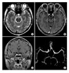
Laboratory findings were as follows: white blood cell count, 2,000/ul; red blood cell count, 3.11×10⁶/ul; hemoglobin, 8.9 mg/dl; platelet count, 246×103/µl; Creactive protein, 4.03 mg/dl. Other serum chemistry values were within normal limits. Brain computed tomography (CT) and CT angiography were normal. Brain magnetic resonance imaging (MRI) with gadolinium enhancement revealed no lesions (Fig. 1). Cerebrospinal fluid (CSF) pressure was 100 mmH2O with protein content of 43 mg/dl and glucose 64 mg/dl. CSF contained no white blood cells and cytologic study showed no abnormal cells.
He received steroid pulse therapy and intrathecal methotrexate therapy in combination with systemic chemotherapy. Headache decreased, but oculomotor nerve palsy remained.
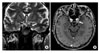
Two weeks later, he visited our department with more aggravated neurologic symptoms and signs. Neurologic examination showed a complete left third nerve palsy with fixed dilated pupil but preserved lateral gaze. Other focal neurologic signs were absent. Follow-up MRI revealed a slightly enhancing mass lesion on the left cavernous sinus which encroached upon the oculomotor nerve (Fig. 2).
He received systemic chemotherapy, but the neurologic sign did not improve. About one month later, he died of disseminated intravascular coagulation (DIC) and upper gastrointestinal bleeding. Postmortem examination was not performed.
Discussion
The most remarkable clinical feature of our case was initially negative findings of brain MRI and CSF cytology and the delayed appearance of mass lesion on cavernous sinus.
The oculomotor nerve branches governing the levator and superior rectus muscles are located in the upper part of the cavernous sinus.2 Initially our patient showed the left eyelid ptosis and vertical gaze palsy, but did not show lateral gaze palsy and pupillary involvement. It was conceivable that the lymphoma invaded the upper branch of oculomotor nerve that controls levator, superior and inferior rectus. However, at that time, we could not find any lesion even with Gadolinium enhanced MRI, which is known to be very effective in demonstrating early involvement of cranial nerve by the lymphoma.3
Complete unilateral oculomotor nerve palsy is usually due the lesion on fascicular or subarachnoid portion of oculomotor nerve. And the oculomotor nerve palsy caused by cavernous sinus lesion is often associated with involvement of the other ocular motor nerves and the ophthalmic branch of the trigeminal nerve.1,4 But, the isolated oculomotor nerve palsy caused by cavernous sinus lesion, such as in our case, is relatively rare.
Lymphomatous involvement of the CNS should be suspected in any patient with lymphoma when suggestive cranial nerve signs develop. The differential diagnosis includes CNS infection, vascular disease and second malignancy. Multiple cranial nerves, particularly the sixth and seventh nerves, are more commonly involved by lymphoma; on the other hand, however, isolated cranial neuropathy is an uncommon presentation.5,6 Rubinstein et al7 reported several cases of isolated cranial neuropathy as the first sign of intracranial metastasis, which included isolated hypoglossal and isolated trigeminal neuropathies. Several cases of isolated oculomotor nerve palsy caused by lymphoma have also been reported.3,8,9 All the previous reports detected mass lesions on brain imaging initially, but in our case we could not detect any lesion on brain MRI initially. Two weeks later, however, when complete oculomotor nerve palsy developed, we could find slightly enhancing mass lesion encroaching on left oculomotor nerve in cavernous sinus.
We advise, therefore, that when isolated oculomotor nerve palsy develops in patients with lymphoma, cavernous sinus involvement should be considered, and even if initial brain imaging and CSF exams reveal negative, follow-up examinations should be performed to detect the cause of neurologic signs in patients with lymphoma.
 XML Download