

 PDF
PDF ePub
ePub Citation
 Print
Print


Introduction
The conventional view of the accessory nerve as being composed of cranial and spinal roots has been challenged by finding the absence of a morphologically distinct cranial root of the accessory nerve.1 In that study, the traditional cranial root of the accessory nerve was suggested to be part of the vagus nerve, and only the spinal root to compose the accessory nerve. More recent study2 has confirmed that the accessory nerve originated form the spinal cord with no distinct cranial contribution, except for one case in which a small, but distinct connection was seen between the vagus and the accessory nerves within the jugular foramen.
The spinal root of the accessory nerve arises as a series of rootlets from the lateral aspect of the first five cervical segments of the spinal cord.3 With regard to the spinal rootlets of the accessory nerve, most anatomy textbooks describe them as descending down to the fifth or sixth cervical segment.4?6 Kumaki (1970),7 however,reported their most caudal level was T1 in Japanese specimens.
We conducted this study to clarify the most caudal level of the spinal rootlets of the accessory nerve on the spinal cord of Koreans, and also to investigate the number of the spinal rootlets at each cervical segment.
Materials and Methods
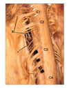
The subjects used in this study were 18 male and 3 female Korean adult cadavers, aged from 20 to 82, with the average of 55 years old. After the calvaria and the dorsal spinal columns were opened, the spinal cords and brain stems were removed en bloc. The dura maters were removed, and forty-two sides of the spinal cords were studied under a surgical microscope. A microscopic hook was frequently used to identify the spinal rootlets of the accessory nerve. The average number of the spinal rootlets was calculated at each cervical segment, and their most caudal level was identified on each specimen.
Results
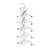
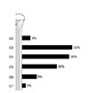
The average number of the spinal rootlets of the accessory nerve was highest at the C1 cervical segment (4.4), followed by 3.1 at the C2, 2.4 at the C3, 0.8 at the C4, and 0.4 at the C5 segment (Fig. 1). One or two rootlets were observed at C6 in 7 cases, and two rootlets were at C7 in only 1 case. The number of the rootlets varied at even the same level of segments depending on the specimens: the maximum being 7 and the minimum being 1 at C1.
Discussion
The present study showed that the most caudal level of the spinal rootlets of the accessory nerve was different depending on the specimens, varying from C2 to C7, and the most frequent level of them was C3,followed by C4, C5, C6, C2, and C7. These results meant the variability in the formation of the accessory nerve, and were in disagreement with those of the previous reports; the most caudal level of the spinal rootlets varied from C2 to T1,7 C3 to C7,8,12 C2 to C6,9,10 and C3 to C5,11 and the most frequent level of them was C3,10 C4,7,9,11,12 C5.8 These differences seemed to be related more with the observation methods and the number of specimens used in each study, rather than the inter-racial variations. That was because the range of the most caudal level of the spinal rootlets was wider in the studies using a stereomicroscope7 and a surgical microscope in this study, than in the studies with the unaided eyes8?11 and a magnifying glass.12 And that was also because the number of specimens was the most in the study7 which showed the widest range of the most caudal level of the spinal rootlets, and the least in the study11 which showed the narrowest range; the number of the specimens used was 104,7 40,8 68,9 38,10 12,11 100,12 and 42 in this study.
This study presented the average number of the spinal rootlets of the accessory nerve at each cervical segment, and demonstrated the higher number of the rootlets at the more cranial level of the cervical segment. The average number of the spinal rootlets in this study was different from a previous one,11 which used the Korean specimens. It was 4.4 and 1.6 at C1,3.1 and 2.3 at C2, 2.4 and 2.2 at C3, 0.8 and 0.9 at C4, 0.4 and 0.3 at C5, in the current and previous studies, respectively. The observation method and the number of specimens might be related with this difference, as in the range of the most caudal level of the spinal rootlets.
In order to understand the formation of the diverse number and range of the spinal rootlets composing the accessory nerves, an embryologic or a comparative anatomical study is required. Considering the diversity
of the spinal rootlets, and the intra- and extradural connections between the accessory and the cervical nerves, there may be differences among the neural routes of the accessory nerves. These possible differences also remain to be clarified.
 XML Download